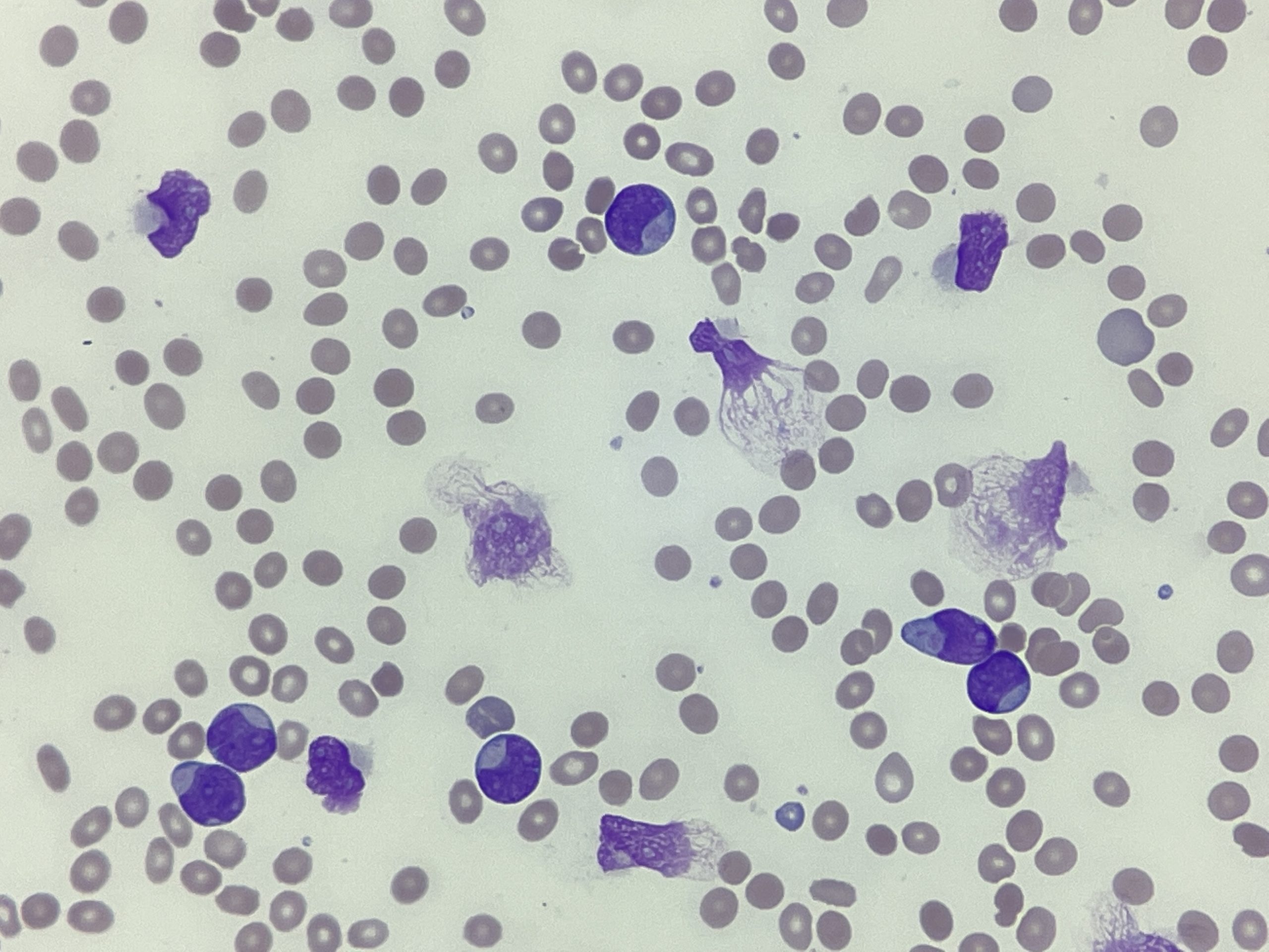
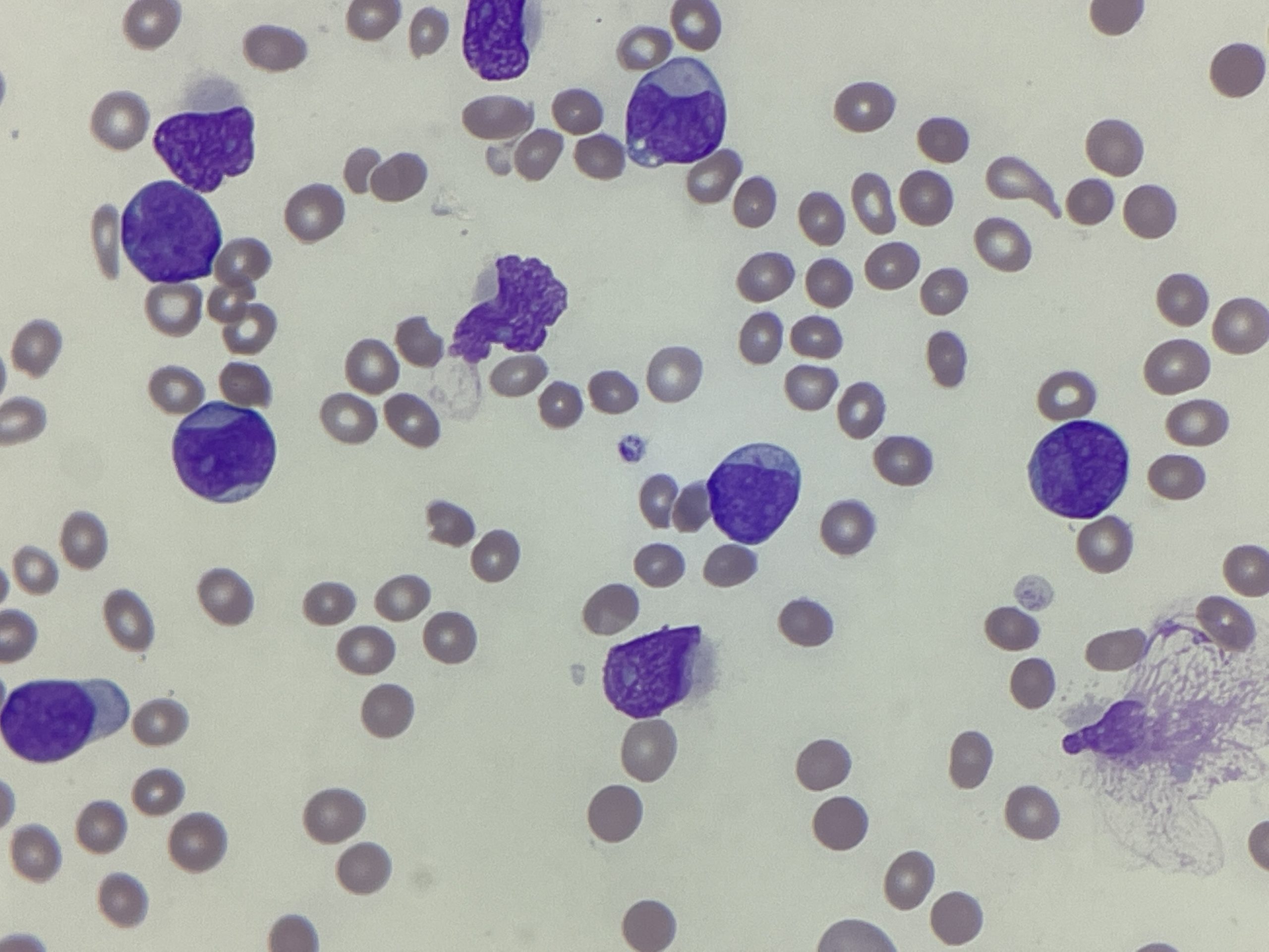
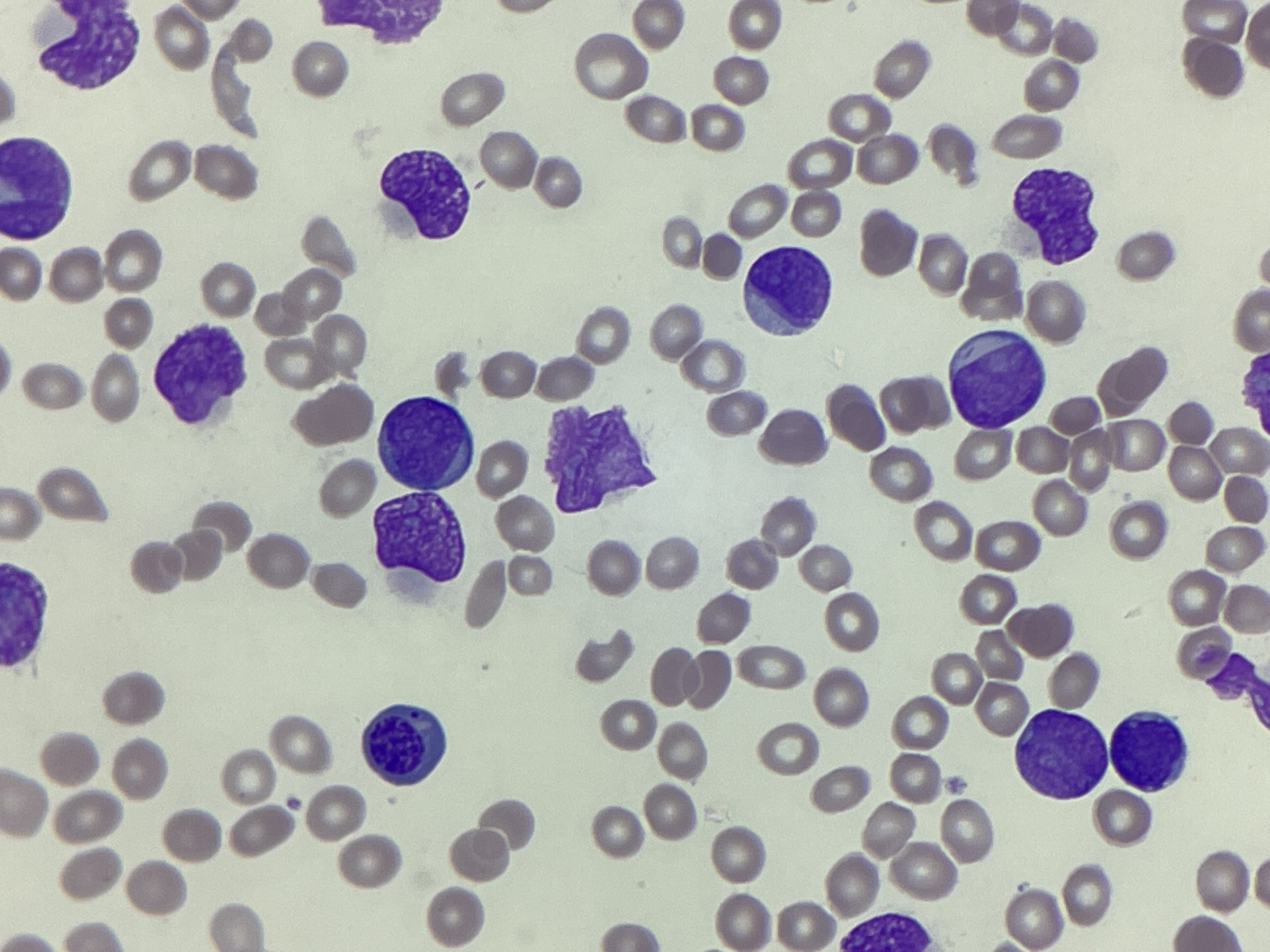
Smear cells

Smear cells (also known as smudge cells) are fragments of cells that have been destroyed during the process of making a blood film. They are usually associated with chronic lymphocytic leukaemia (CLL), but that is not the only condition they are seen in. Cells smear due to fragility of the membrane. In the image above and those below, there are numerous smear cells. Looking at the surrounding cells however, predominantly blasts can be seen. 98% blasts were counted in a manual differential count. Some of the blasts have a cup like / fish mouth appearance and others contain Auer rods. The cup like shape of the nuclei in the blasts is indicative of the NPM1/FLT3 mutation.


Similar to the smear cells, Auer rods are the same. Just because they’re seen in AML as in the images above, doesn’t mean they are confirmatory for it. They have also been reported in neutrophils and lymphoblasts as well!
_____
Images: Personal photography.
Privacy Policy | Refund & Return Policy | Only Cells LTD © 2025
