P. falciparum

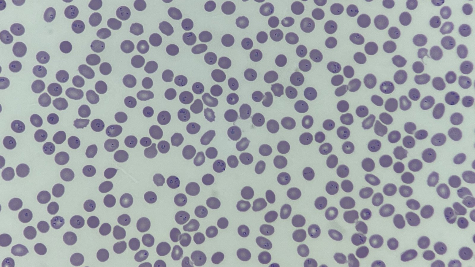
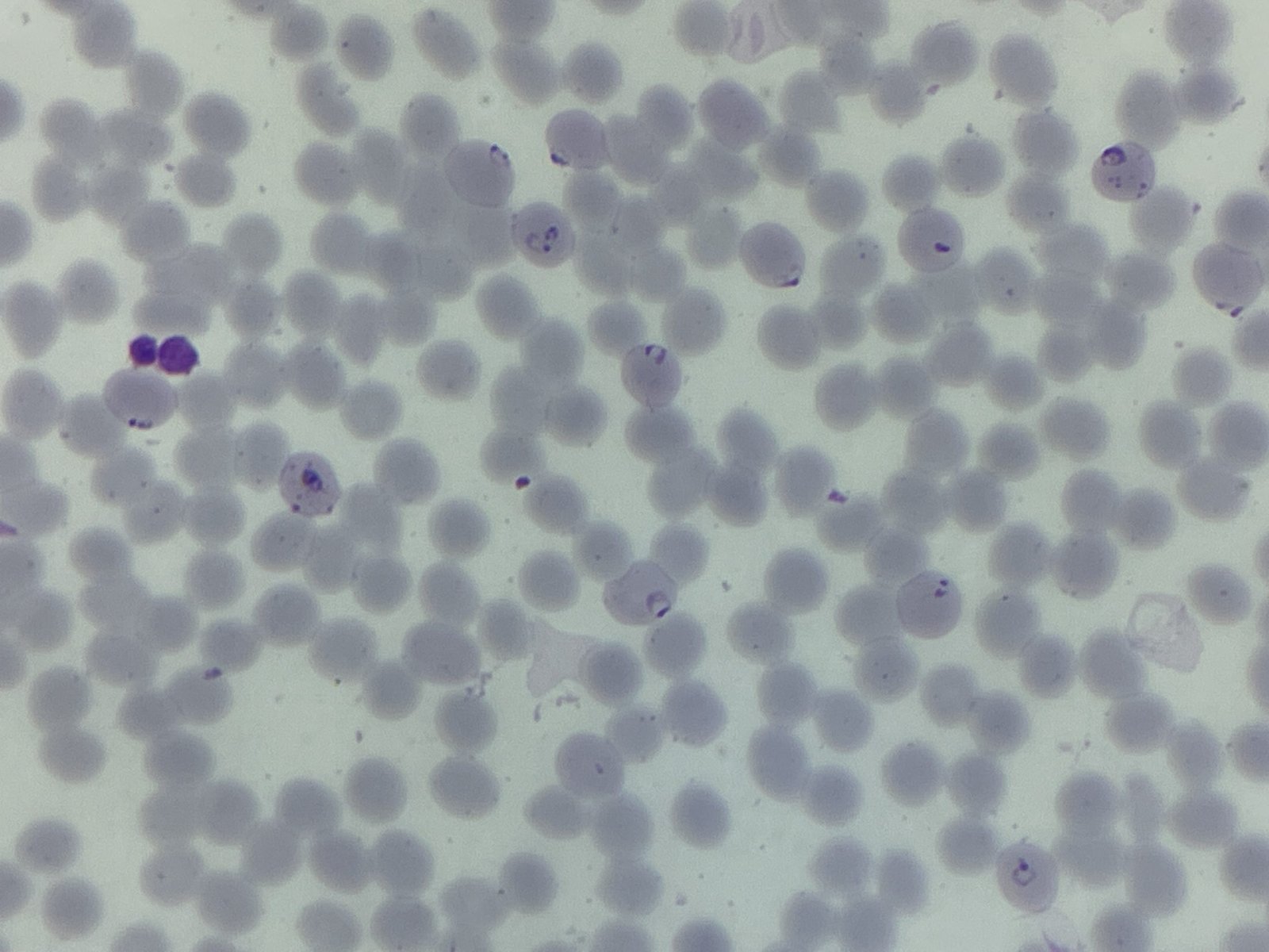
The blood film image shows small, fine ring parasites which in some cases have double chromatin dots. The infected red cells are similar in colour to those not infected and there seems to be no colour change either.

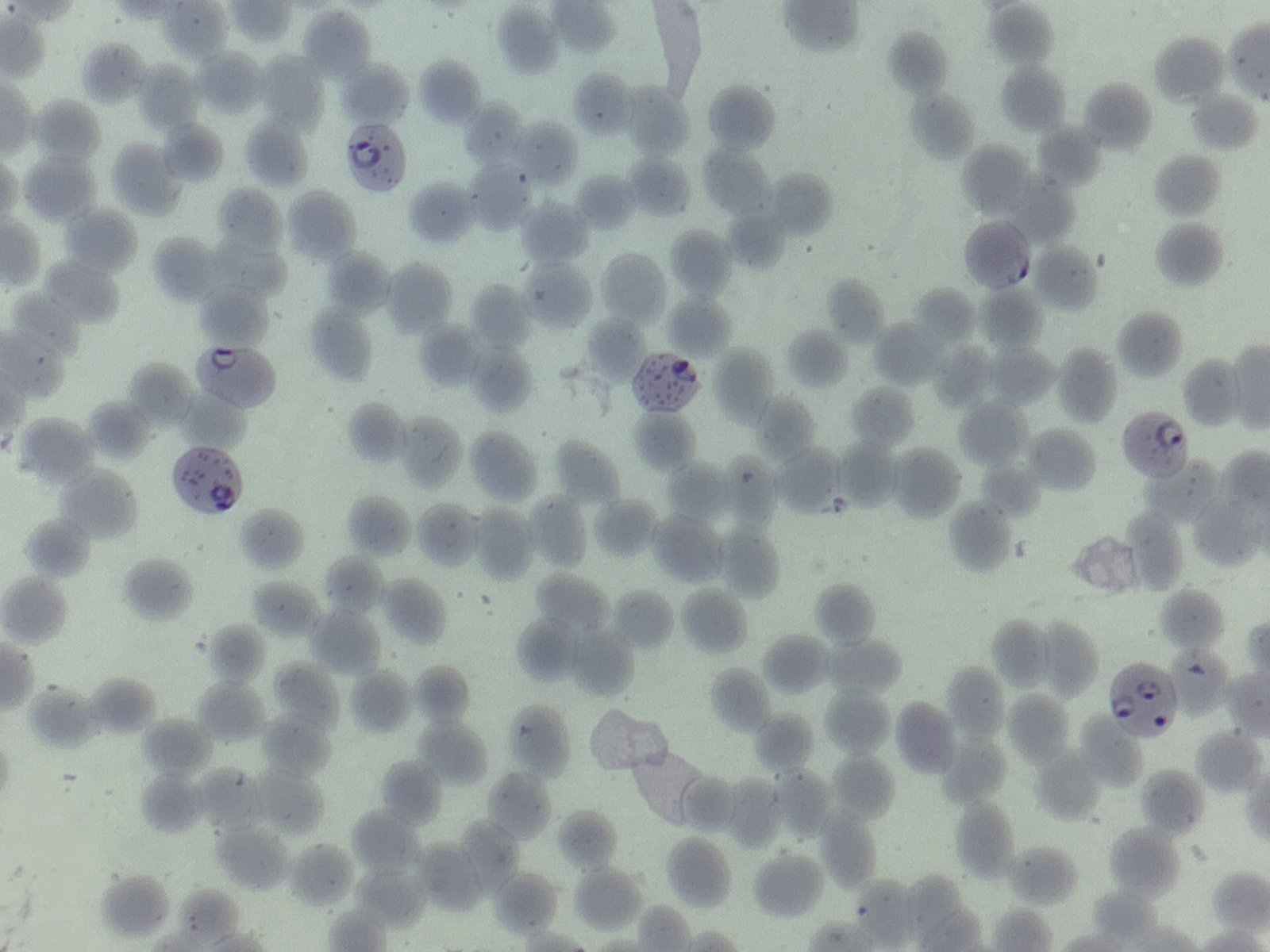
There are accole forms. These are ring forms of the parasite that generate a ‘blister’ on the edge of the cell and is characteristic of a P. falciparum infection. Some of the red cells contain multiple parasites.

The infected red cells contain Maurer’s clefts, membranous structures, which are seen in a falciparum infection. One way to distinguish the various inclusions seen in the red cells during infection by the different Plasmodium species, such as James’ and Schüffner’s dots from the Maurer’s clefts seen in P. falciparum is that clefts can be counted a lot more easily than the dots!
_____
Images: Personal photography.
Privacy Policy | Refund & Return Policy | Only Cells LTD © 2025
