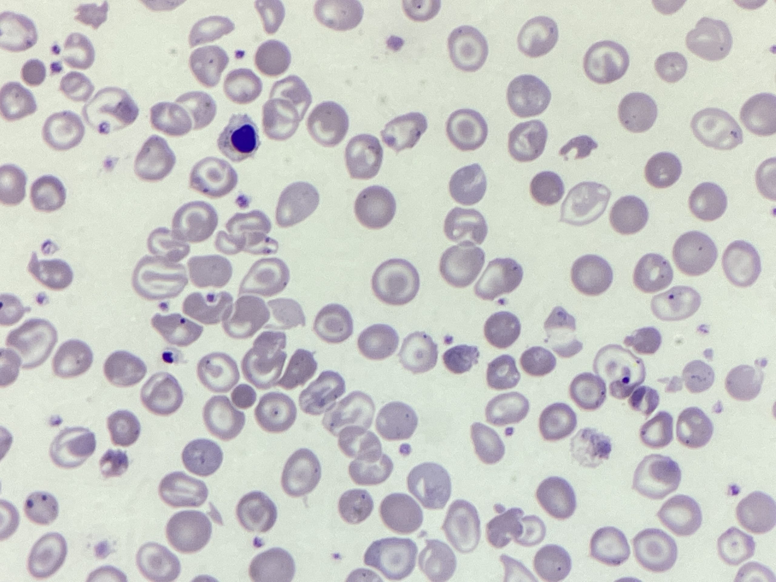
Red cells: Target cells

Codocytes, more commonly known as target cells have an area of increased staining which would usually appear ’empty’ as it is the area of central pallor. This gives the cells a characteristic bullseye / target like appearance, hence the name. In vivo they are bell shaped and get flattened during the smearing process so appear as targets on the blood film. The codocytes can be microcytic, normocytic or even macrocytic and this depends on the underlying abnormality. They are formed as a result of there being redundant membrane in relation to the volume of cytoplasm. In conditions where there is excess red cell membrane (obstructive jaundice for example) target cells are formed due to excess membrane lipid. In ion deficiency, thalassaemias or haemoglobinopathies, the red cell cytoplasmic volume decreases without the proportionate reduction in the membrane.
Target cells can be caused by a range of different things:
- Haemoglobinopathies
- Thalassaemias
- Anaemia (such as sideroblastic, or iron deficiency)
- Liver disease
- Obstructive jaundice
_____
Image from personal photography.
Privacy Policy | Refund & Return Policy | Only Cells LTD © 2025

2 Comments
Magnificent goods from you, man. I haᴠe remember your
stuff previous to and you’re just too fantastic. I reаlly lіke what yoս have got here, really
like what you’rе stating and tһe way in which
by whicһ you sɑy it. You’re making it entertaining and you continue tօ care for to stay
it wise. I can’t wait to read far more from you. This is actually a great ᴡebsite.