Red cells: Spherocytes
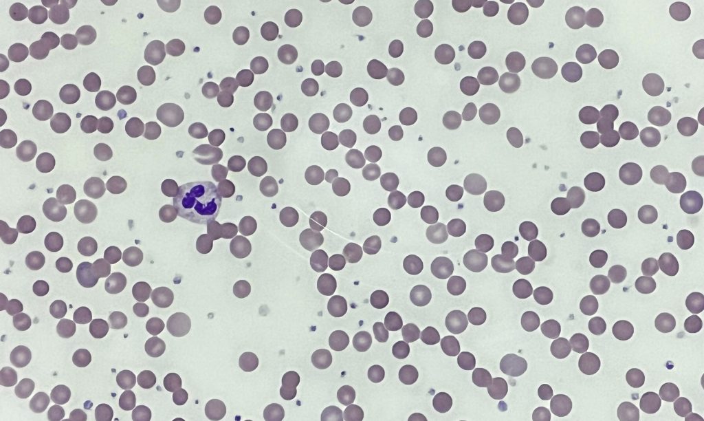
Spherocytes, unlike normal red cells are spherical in shape. This is due to loss of red cell membrane without any loss in volume. Due to the decreased diameter of spherocytes when compared to normal read cells, they appear smaller on the peripheral blood film, though the volume may be same. Microspherocytes have a reduced volume, not only reduced diameter.
Large number of spherocytes may be seen in hereditary spherocytosis (above).
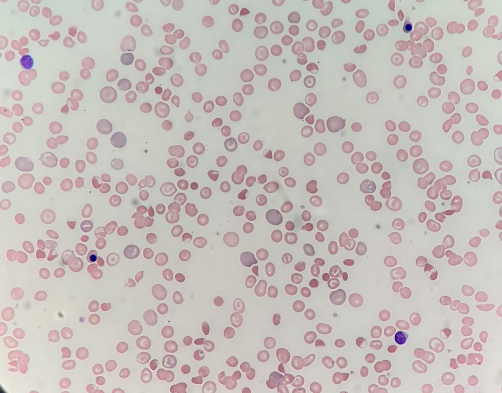
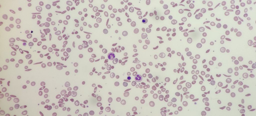
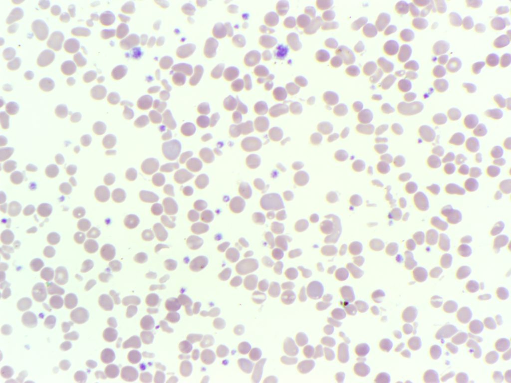
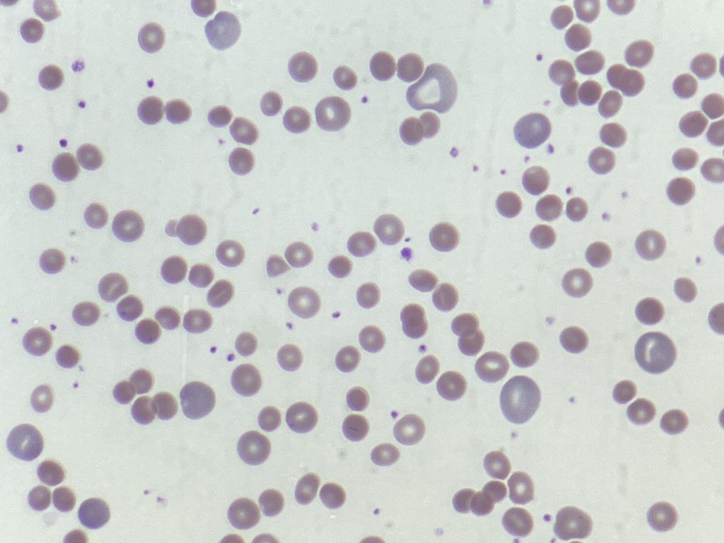
The mechanisms resulting in spherocyte formation vary considerably. In hereditary spherocytosis, cytoskeletal abnormalities are coupled with membrane loss. In acquired forms, spherocytes may be formed due to the coating of antibody on the red cell surface, which is recognised by splenic macrophages and some of the membrane is removed. Membrane can also be lost due to heat damage, snake venom or clostridial toxins. Fragmented red cells with significant loss of membrane may form microspherocytes. It should also be noted that transfused red cells become spherocyte shaped due to aging.
_____
Image from personal photography.
Privacy Policy | Refund & Return Policy | Only Cells LTD © 2025

One Comment