Customize Consent Preferences

We use cookies to help you navigate efficiently and perform certain functions. You will find detailed information about all cookies under each consent category below.

The cookies that are categorized as "Necessary" are stored on your browser as they are essential for enabling the basic functionalities of the site. ... 

Always Active

Necessary cookies are required to enable the basic features of this site, such as providing secure log-in or adjusting your consent preferences. These cookies do not store any personally identifiable data.

No cookies to display.

Functional cookies help perform certain functionalities like sharing the content of the website on social media platforms, collecting feedback, and other third-party features.

No cookies to display.

Analytical cookies are used to understand how visitors interact with the website. These cookies help provide information on metrics such as the number of visitors, bounce rate, traffic source, etc.

No cookies to display.

Performance cookies are used to understand and analyze the key performance indexes of the website which helps in delivering a better user experience for the visitors.

No cookies to display.

Advertisement cookies are used to provide visitors with customized advertisements based on the pages you visited previously and to analyze the effectiveness of the ad campaigns.

No cookies to display.

🤍

🤍

Red cells: Basophilic stippling
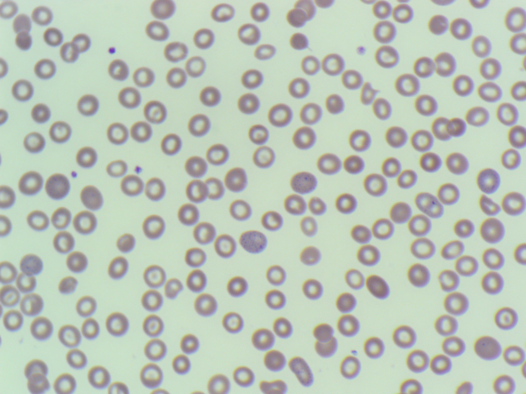
Blood film showing basophilic stippling (2 cells at centre and on near 8 o’clock).
Basophilic stippling in a patient with aplastic anaemia.

Basophilic stippling refers to the presence of evenly distributed inclusions within the erythrocytes. They are formed from aggregates of ribosomes and degenerating mitochondria. They mainly consist of RNA. In basophilic stippling the granules are evenly distributed throughout the cell which helps to differentiate them from other inclusions such as Howell-Jolly Bodies (single, circular inclusion) and pappenheimer bodies (small cluster at the periphery of the cell). They are seen a range of different conditions:

  • β-thalassaemia minor
  • Thalassaemia major
  • Haemolytic anaemia
  • Sideroblastic anaemia
  • Liver disease
  • Heavy metal poisoning (lead, arsenic, zinc, silver, mercury)
  • Deficiency of pyrimidine 5-nucleotidase

_____
Image from personal photography.

Leave a Reply

Your email address will not be published. Required fields are marked *