This week’s Morphology Monday featured a 46-year-old male who presented with fatigue, mild shortness of breath, and light-headedness. His Full Blood Count showed the following:
FBC Results:
- Hb: 104 g/L
- RBC: 3.5 × 10¹²/L
- HCT: 0.32
- MCV: 91 fL
- MCH: 29.7 pg
- MCHC: 326 g/L
- RDW: 14.2%
- WBC: 6.7 × 10⁹/L
- Platelets: 480 × 10⁹/L
A blood film was prepared for review, and the key morphological feature for participants to identify was a stomatocyte.
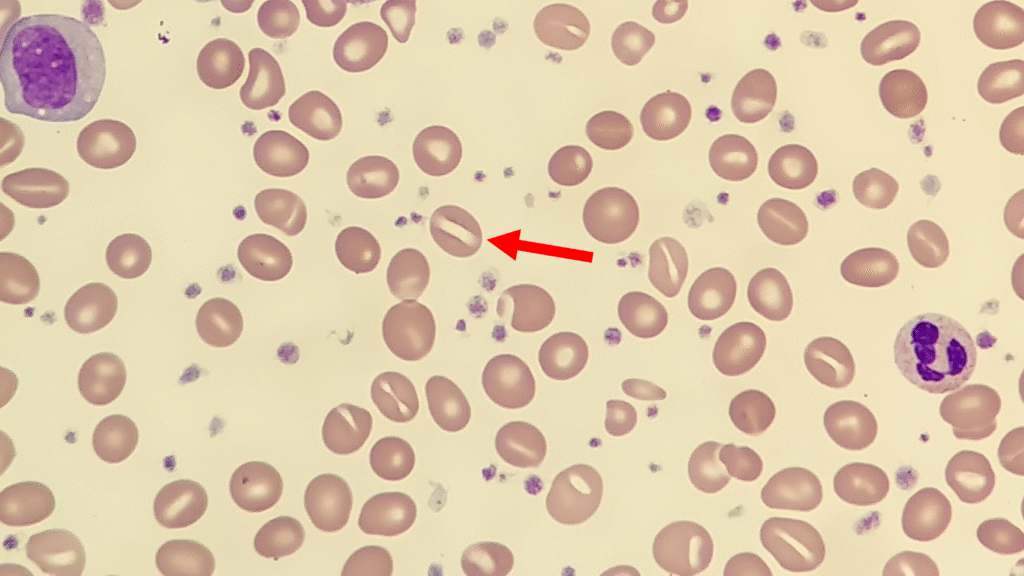
The central pallor of stomatocytes appears as a slit- or mouth-shaped area, rather than the typical circular central pallor of normal red cells. They may be present in small numbers without clinical significance, but when increased, they can point towards underlying disorders.
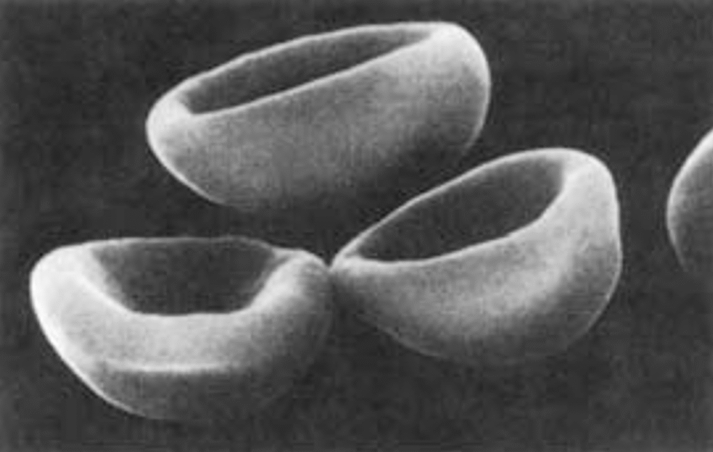
Interestingly, in vivo stomatocytes are cup or bowl shaped which can be seen from the scanning electron microscopic image below1:
Stomatocytes can be found in a range of clinical conditions, including:
Hereditary causes:
- Hereditary stomatocytosis (overhydrated and dehydrated variants) – rare congenital membrane disorders characterised by altered red cell permeability and haemolytic anaemia.
Acquired causes:
- Liver disease, particularly alcoholic liver disease
- Alcohol excess (may resolve with abstinence)
- Medications, such as certain chemotherapeutic or psychotropic agents
- Rh-null phenotype (rare)
- Acute infections or transient red cell membrane stress
- Artifact due to suboptimal smear preparation or excess EDTA exposure
_____
1 Image from Cells, Bain B. Fourth Edition
2 Comments
What was the cause of the stomatocytosis in this case?
Thanks for popping by and for the comment Louise. This was an old image I took many years ago and did not at the time record the cause, so unfortunately say in this case.