Earlier in the week a couple of blood film images were shared as part of this week’s Morphology Monday case. On closer inspection the blood film revealed both trophozoites and schizonts of Plasmodium malariae.
The first thing to note is the infected red cells are normal or slightly smaller in size and do not contain any stippling.
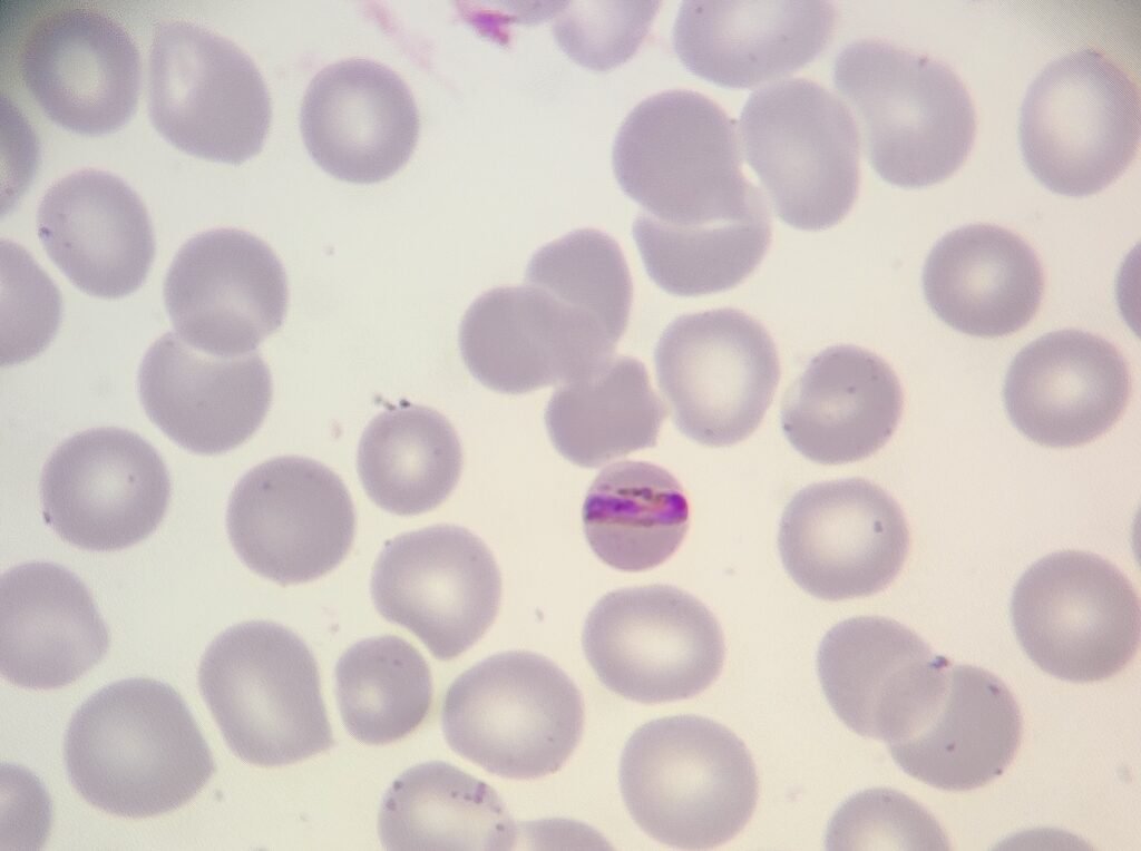
The trophozoite is neat and compact, stretching across the cell in a distinct band form.
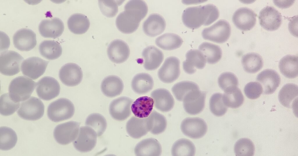
The schizont on the other hand are striking. They contain up to 12 nuclei (I can count 11 in the image above!) and densely packed pigment that give them a golden brown hue.
Both these features are classic for P. malariae, one of the less common causes of malaria but with characteristic morphology that sets it apart from other Plasmodium species.