Happy New Year, everyone! We are kicking off the first Morphology Monday of the year with a case that highlights the importance of “eyes-on” microscopy versus automated flagging.
The context:
- 62 year old female.
- Presented to Emergency Department overnight
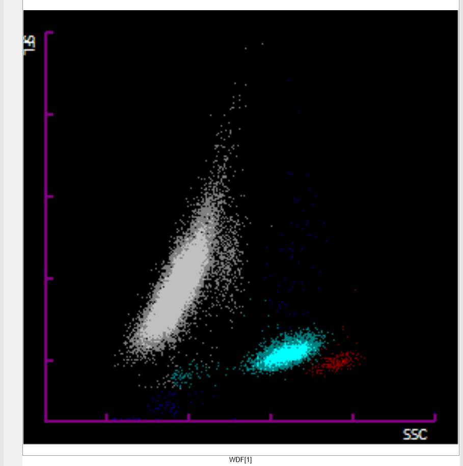
- The analyser gave a grey population when doing the differential (see below).
Laboratory findings (initial):
The FBC showed:
- WBC: 38.39 x10⁹
- Haemoglobin: 112g/L
- MCV: 78.4 fL
The blood film was made to perform a manual differential count. The “grey zone” on a scatterplot often indicates cells that the analyser cannot confidently categorise, frequently seen when cells have unusual size or internal complexity.
The biomedical scientist working overnight reported a large percentage of blast cells. When the film was reviewed the following day the following was seen:
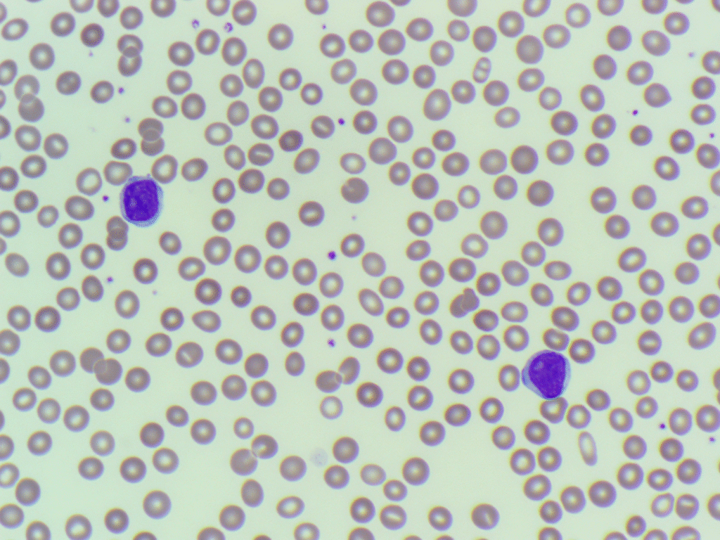
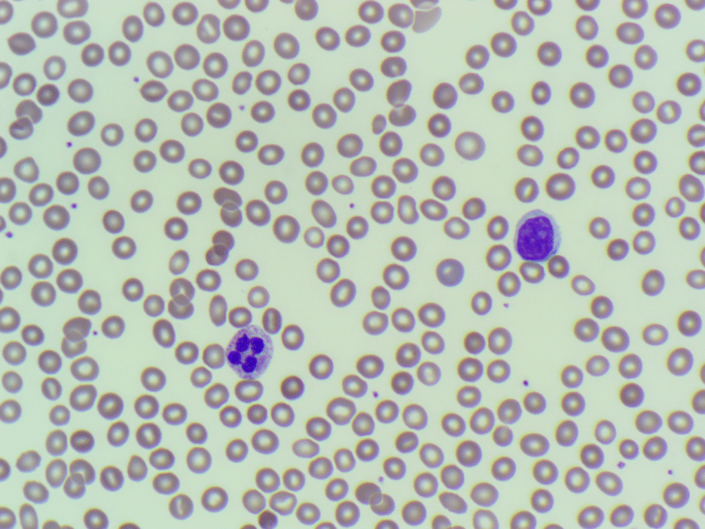
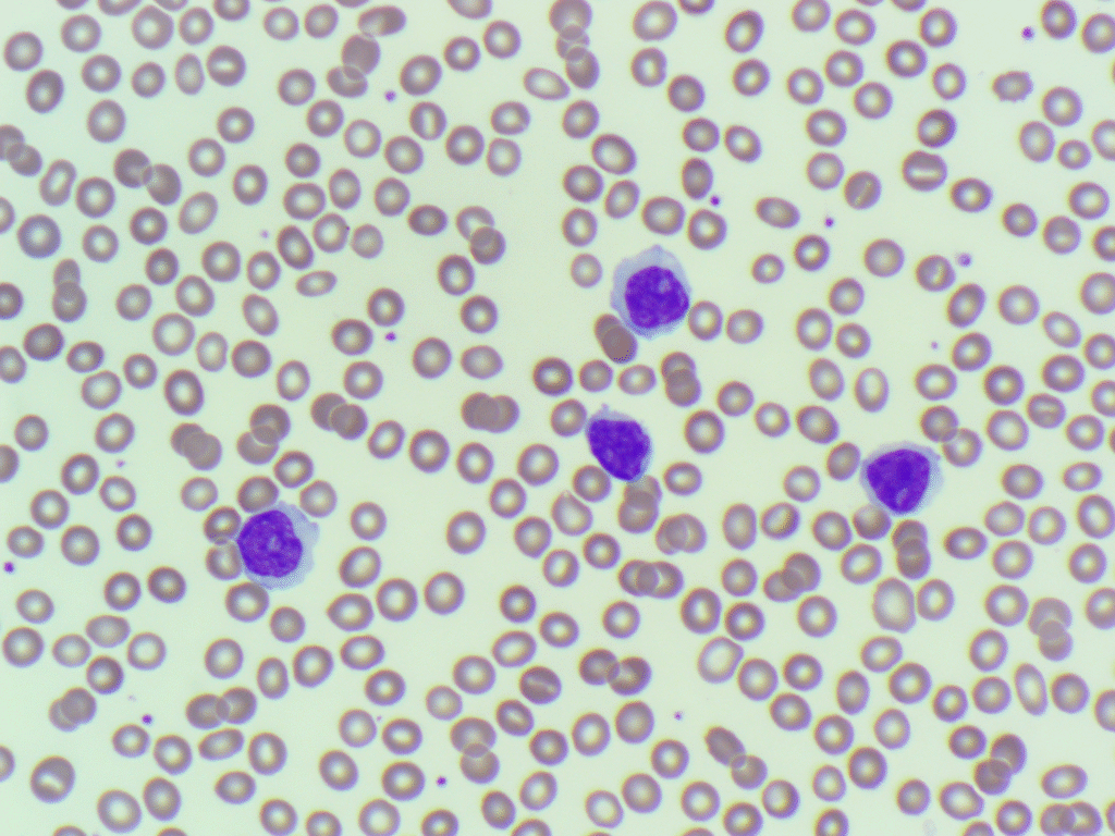
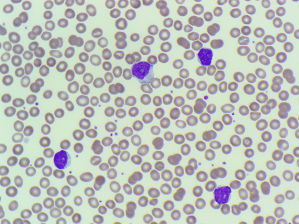
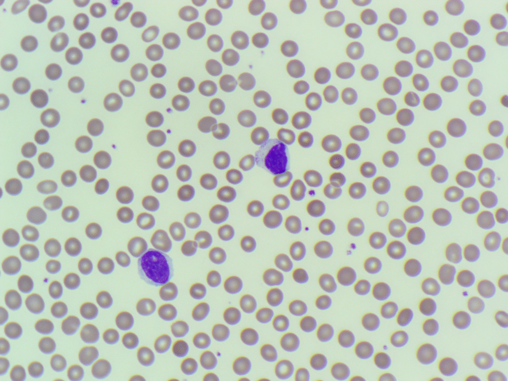
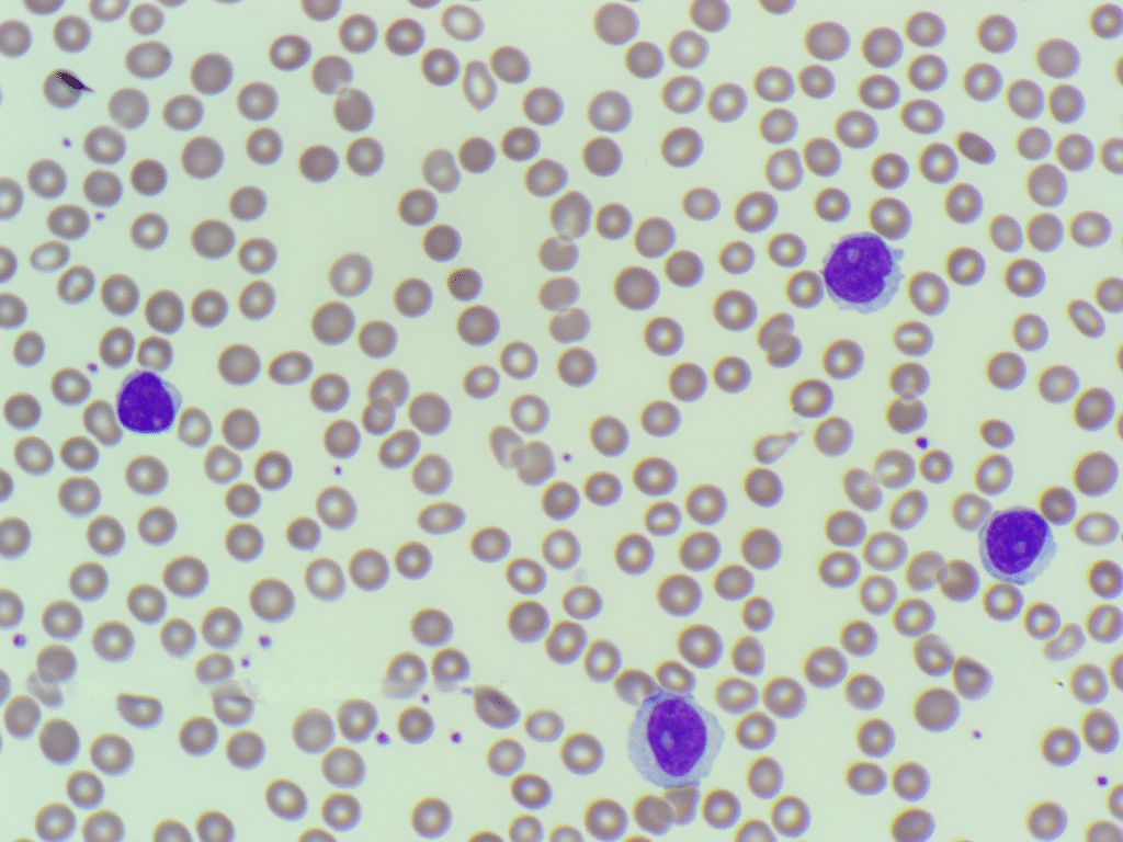

Take a close look at the scattergram and the attached images.
- Do you agree with the initial assessment of these cells as “Blasts”?
- If they aren’t blasts, what morphological features might have led the initial scientist to that conclusion?
- Based on the patient’s age and the provided data, what is your top differential diagnosis?
Post your thoughts and film descriptions in the comments! We will reveal the final diagnosis and the teaching points later this week. You can also follow the case on LinkedIn and X (Twitter).
One Comment
Prolymphs maybe