Thank you to everyone who weighed in on this week’s case! It was a fantastic example of how a single blood film can tell a complex story of genetics, clinical intervention, and surgical history.
The patient is missing their spleen. In addition to the underlying HbE/β-thalassaemia, the film showed classic “post-splenectomy” features that occur when the body’s primary “quality control” filter is removed.
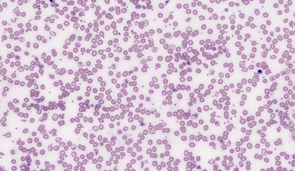
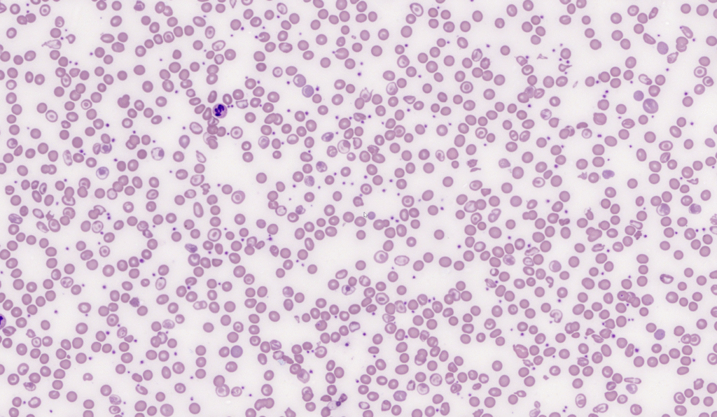
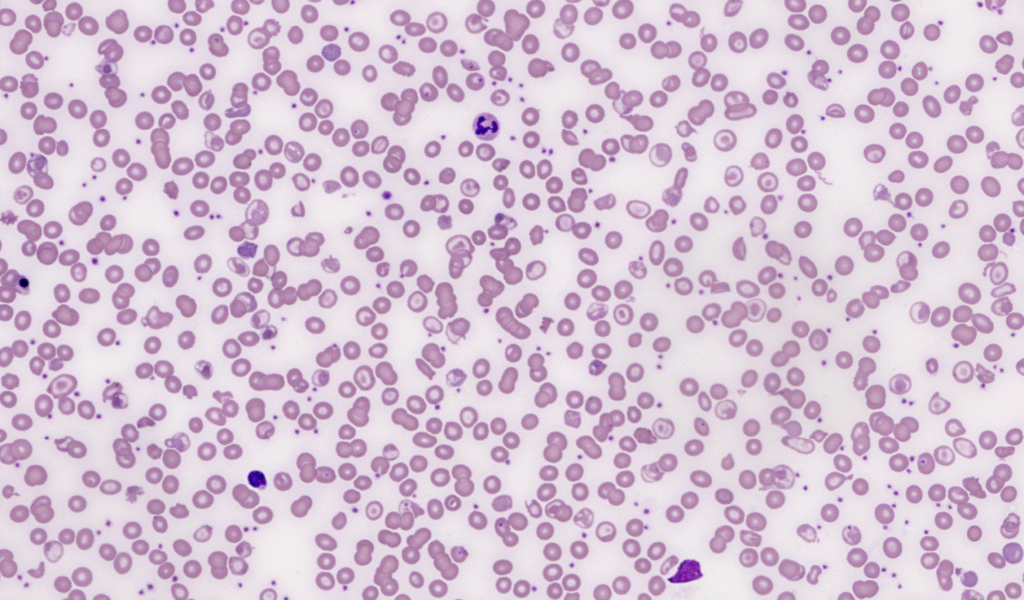
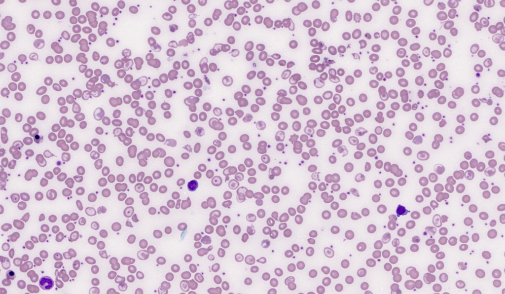
Because this patient is transfusion-dependent, the film displays two distinct populations of red cells (a dimorphic picture):
- Donor Cells: Normochromic and normocytic (MCV and MCHC within reference ranges). These are the healthy “standard” cells from the transfusion.
- Native Cells: The patient’s own cells, which are severely microcytic (small) and hypochromic (pale). This is due to the defective haemoglobin synthesis inherent in HbE/beta-thalassaemia.
When the spleen is removed (often but not always done in thalassaemia to reduce red cell sequestration and transfusion requirements), several features persist in the peripheral blood that would normally be “pitted” out by splenic macrophages:
- Howell-Jolly Bodies: Small, round, purple DNA nuclear remnants. Normally, the spleen removes these “pips” from young red cells.
- Target Cells (Codocytes): These are prominent due to an increase in the surface-area-to-volume ratio. In this case, both the thalassaemia itself and the lack of a spleen (which normally grooms excess red cell membrane) contribute to their presence.
- Pappenheimer Bodies: Siderotic (iron) granules that appear as small blue inclusions, often in clusters, which the spleen can no longer clear.
As one user mentioned, “to be fair the shortlist of organs you can do without is pretty small.”