Thank you to everyone who weighed in on this week’s tropical haematology mystery. This case highlights how a patient’s travel history is an important important diagnostic tool in the laboratory.
The patient is a 34 year old aid worker who recently returned to the UK after two years in the rainforests of Cameroon and Gabon. They presented with recurrent, itchy, non-pitting oedema on the wrists and a “creeping” sensation under the skin. The eosinophilia (4.2 x10⁹/L) coupled with a decreased platelet count prompted the making of a blood film.
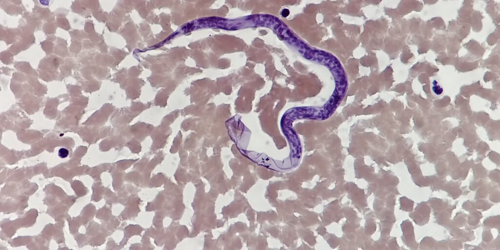
The parasite identified is Loa loa, a filarial nematode endemic to West and Central Africa. It is transmitted to humans via the bite of an infected Chrysops fly (deer fly). Certain morphological features can help distinguish Loa loa from other microfilariae:
The “sheath” test:
- Loa loa is a sheathed microfilaria.
- On a Giemsa-stained film, the sheath may appear as a delicate, transparent membrane extending beyond the head and tail, though it sometimes stains poorly and requires careful adjustment of the microscope’s fine focus.
The Tail Nuclei: “Lower and Lower”:
- In Loa loa, the nuclei are coarse and crowded, extending in a solid, irregular row right to the very tip of the tail.
- You can remember this by noting that the nuclei don’t stop early; they continue “lower and lower” until they fill the entire caudal extremity.
- In W. bancrofti (another sheathed microfilaria), the tail is pointed and contains no nuclei at the tip.
- Contrast with Brugia malayi, In B. malayi, the nuclei extend toward the tail but are interrupted, typically leaving two distinct, terminal nuclei separated from the main column.