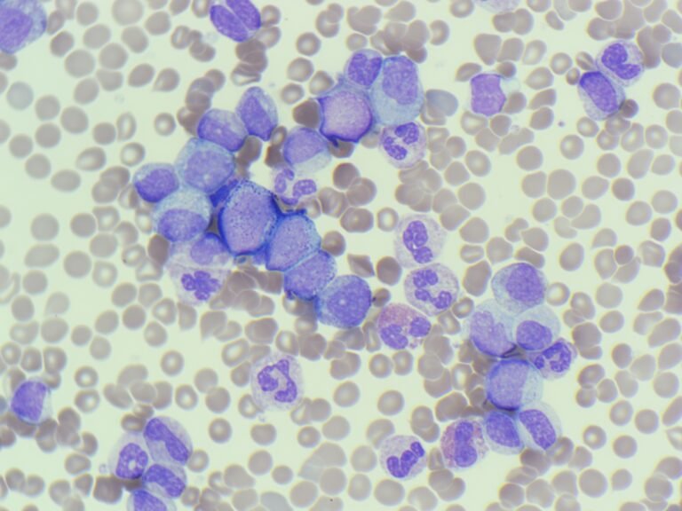
MM260126: CML
This week’s case started with a “heads-up” call from the Haematology SpR before the sample even reached the lab. The staggering WBC and the specific clinical request made for an interesting discussion.

This week’s case started with a “heads-up” call from the Haematology SpR before the sample even reached the lab. The staggering WBC and the specific clinical request made for an interesting discussion.
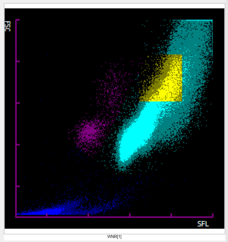
This week’s case began with an urgent call from the haematology Specialist Registrar (SpR) before the sample had even reached the laboratory. A patient had been referred from another hospital and presented to the Emergency Department with a massive clinical picture.
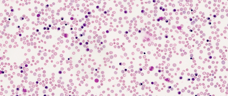
Based on these findings, the diagnosis is Haemolytic Disease of the Foetus and Newborn (HDFN) caused by Rh sensitisation, following the clinical scenario where the mother did not receive anti-D prophylaxis.
This week’s case comes from the Neonatal Intensive Care Unit. A newborn has presented with early-onset jaundice and a rapidly falling haemoglobin level.
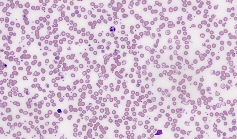
The patient is missing their spleen. In addition to the underlying HbE/β-thalassaemia, the film showed classic “post-splenectomy” features that occur when the body’s primary “quality control” filter is removed.
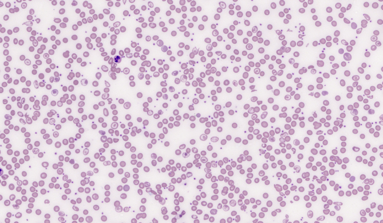
The blood film images are from a transfusion dependent patient who is a compound heterozygote for HbE/beta-thalassaemia.
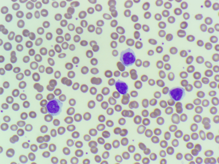
Don’t let a nucleolus default you to “Blast”: Always assess the chromatin density.
62 year old patient presented to ED. Her FBC showed an increased white cell population with the analyser unable to differentiate them properly. The BMS reported them as blasts. Was this correct?
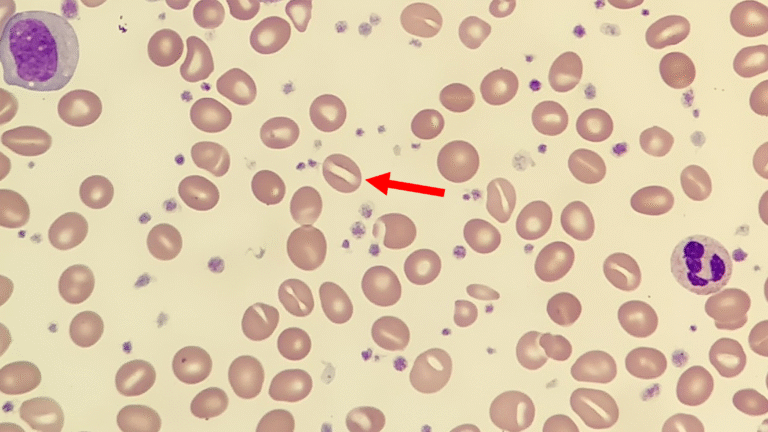
A blood film was prepared for review, and the key morphological feature for participants to identify was a stomatocyte.
This week’s case comes from a 46 year old male who presented to his GP with no significant past medical history, does not take regular medications, and reports no recent infections. He drinks alcohol socially but not excessively.