Morphology Monday | Case MM251013
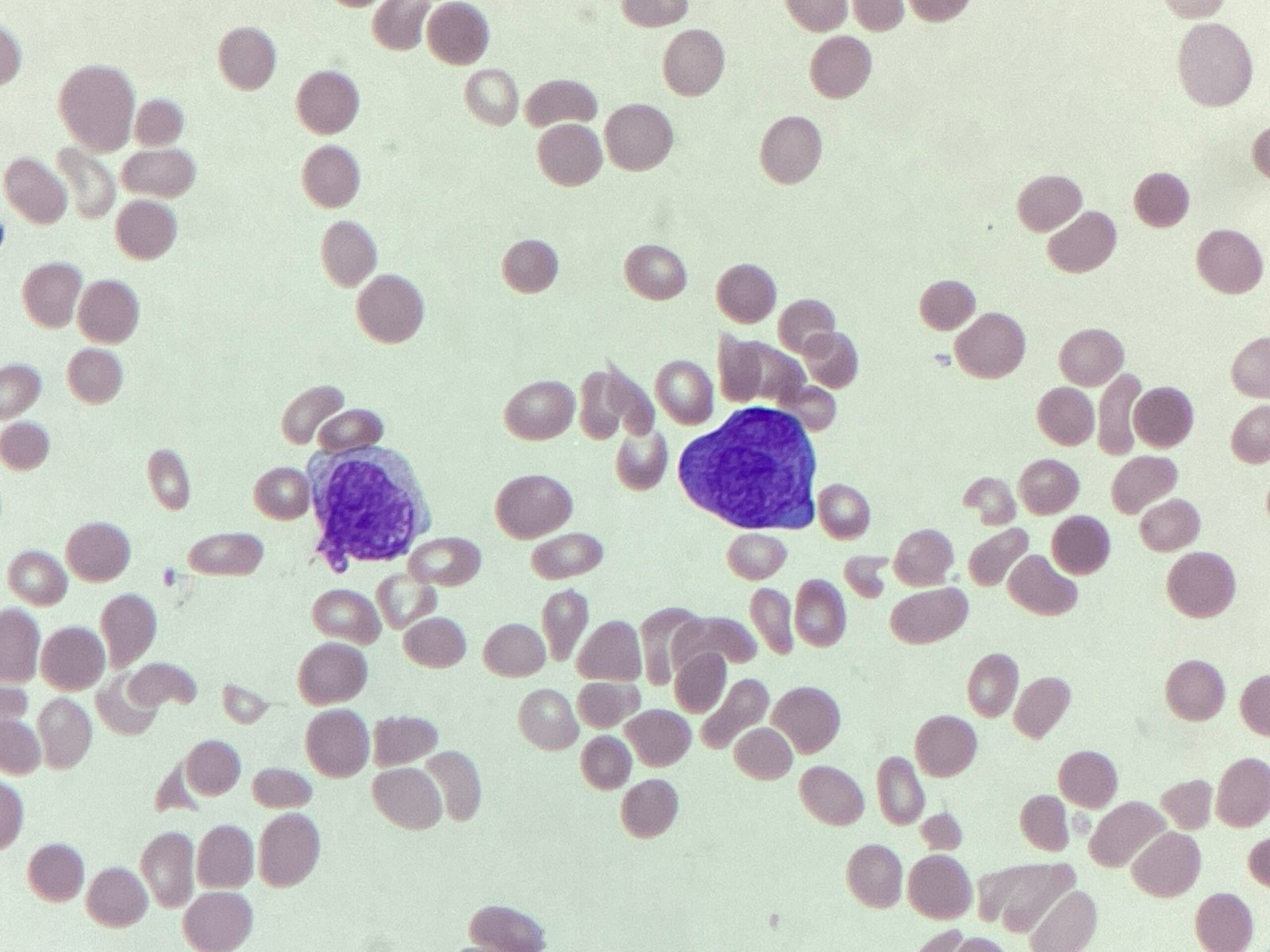
This week’s Morphology Monday case is from a 57 year old who presented to the emergency department.
This week’s Morphology Monday case is from a 57 year old who presented to the emergency department.
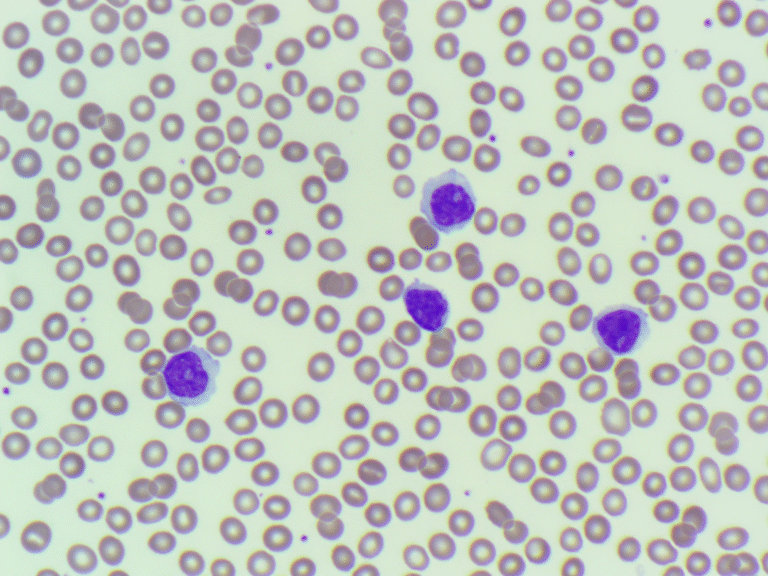
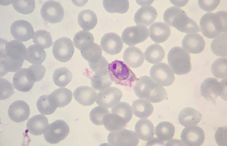
The findings are in keeping with a lymphoproliferative disorder (LPD). Given the predominance of prolymphocytes, the picture is suggestive of Prolymphocytic Leukaemia (PLL). However, this would require confirmation by flow cytometry to establish lineage and clonality.
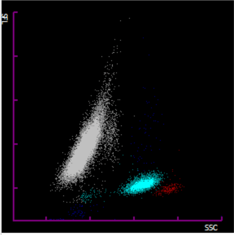
This week’s case comes from a 62-year-old female whose sample was flagged by the analyser, but interestingly, no differential count was produced. The WBC scattergram showed an abnormal grey plot, prompting a manual film review.
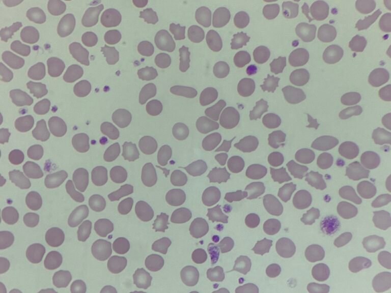
The case underlines how multiple comorbidities can converge to produce complex red cell morphology.
This week’s case comes from a patient with a complex clinical background. The patient presented with anaemia and a raised platelet count, alongside signs of systemic illness.
The blood film, stained with a supravital stain, revealed distinctive “golf ball” inclusions, blue precipitates within red cells.
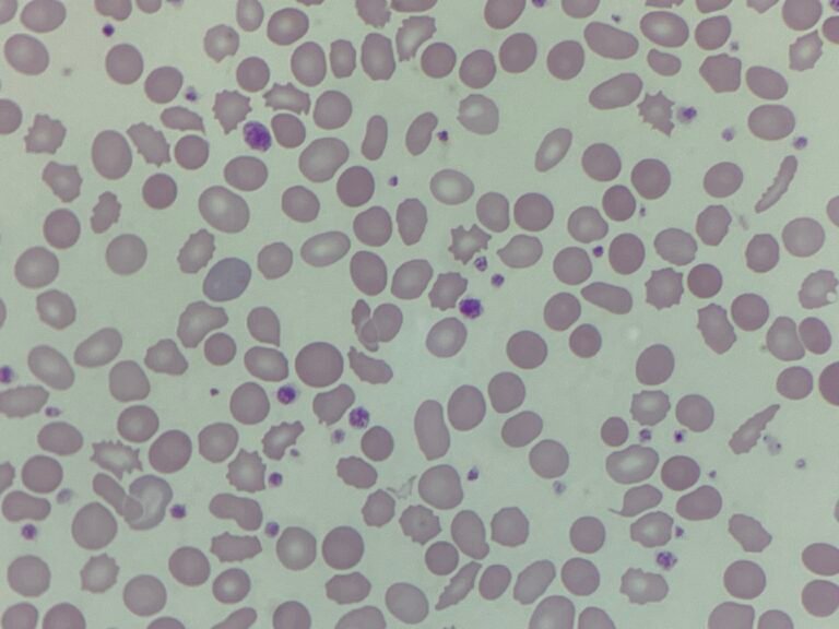
This week’s case comes from a 35-year-old patient who presented with fatigue, mild shortness of breath and intermittent anaemia
The overall findings were consistent with Plasmodium ovale infection, one of the less common species of malaria. It is typically associated with relapsing infections due to dormant liver hypnozoites and is often distinguished by oval-shaped infected red cells with fimbriated edges.
![ABID – The weird and the wonderful webinar [3/3]](https://onlycells.co.uk/wp-content/uploads/ABID-3_Thumbnail-768x432.jpg)
Following on from the foundations laid in Session 1: Tools of the Trade, and applications of those key concepts in Session 2: Putting it into practice, this session focused on some of the more complex techniques used and cases.
This week’s case comes from a 29-year-old male who returned from a recent trip abroad. He presented to the emergency department with fever, chills, sweats, headache, and general malaise.