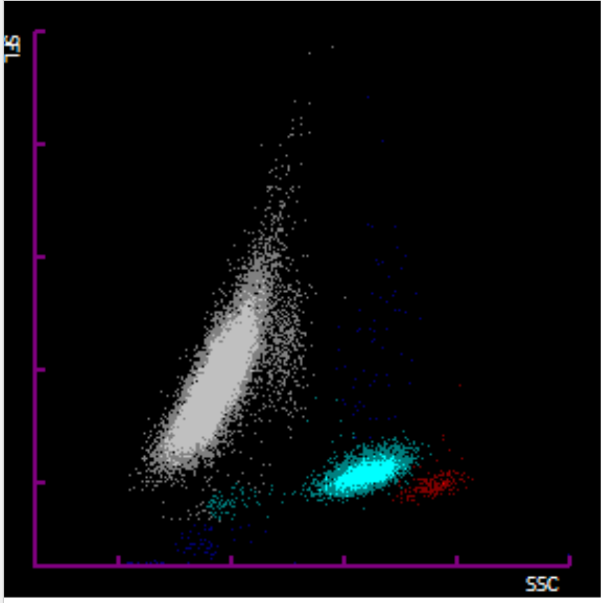
This week’s case involved a 62-year-old female whose full blood count showed a marked lymphocytosis (WBC 34 × 10⁹/L, lymphocytes 90%). The analyser failed to provide a differential, displaying a grey scatter plot, prompting a manual film review.
The film revealed numerous lymphoid cells with prominent nucleoli, consistent with prolymphocytes. Red cells showed mild anisocytosis, and platelets appeared normal.
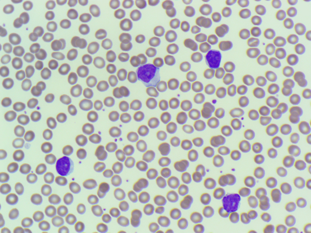
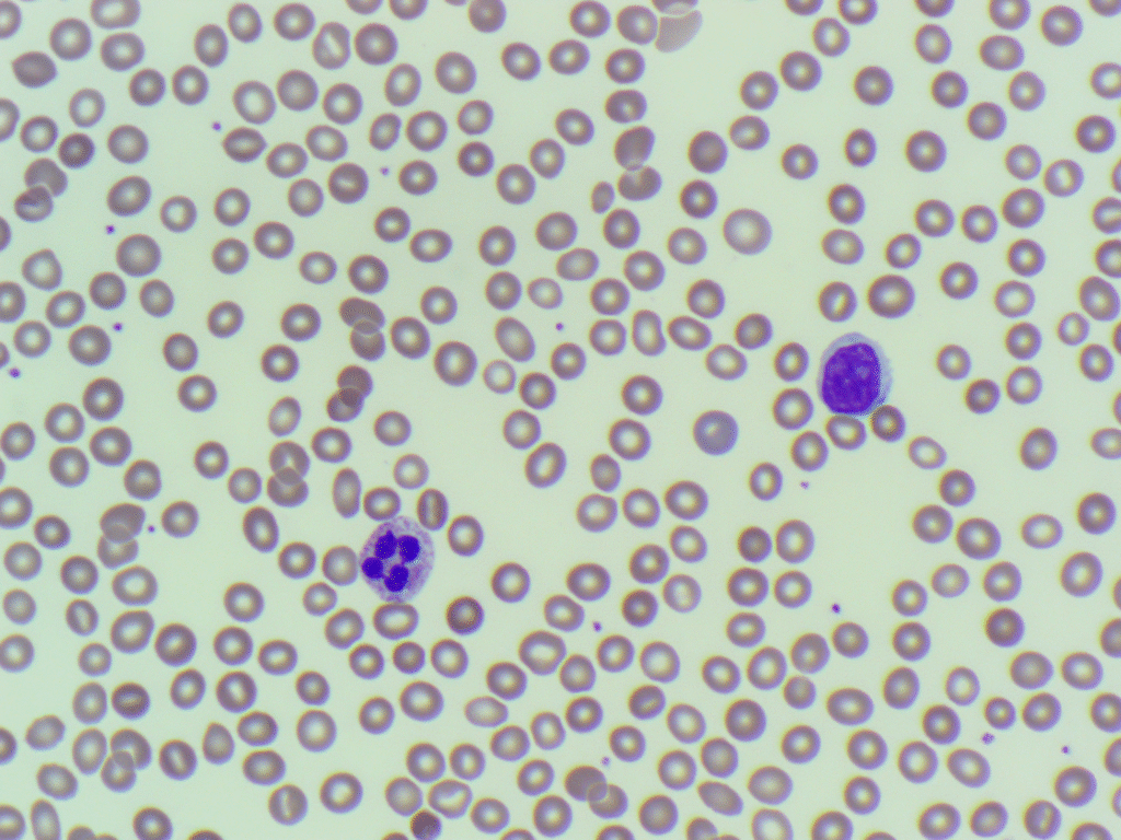
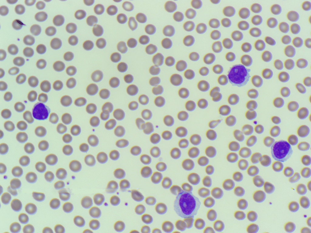
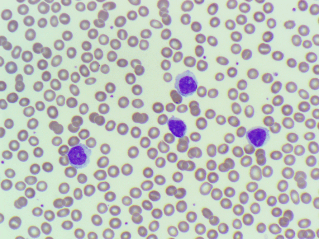
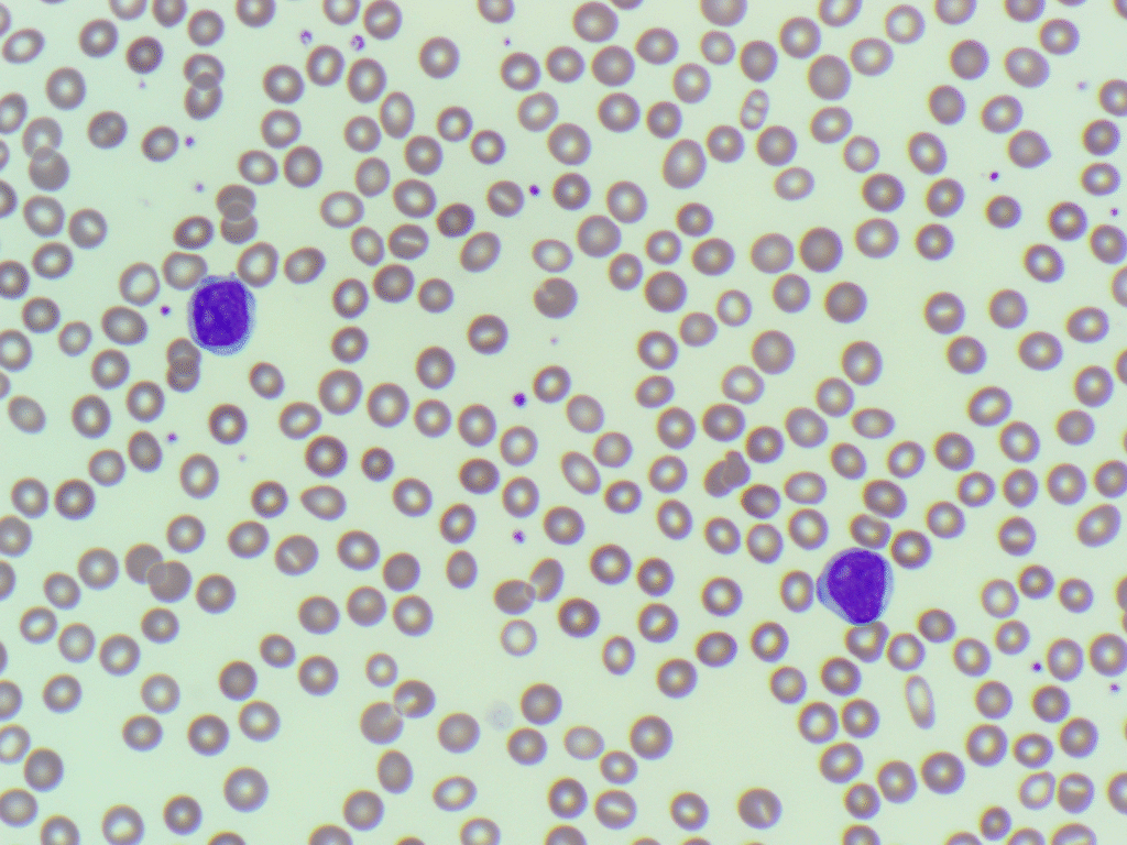
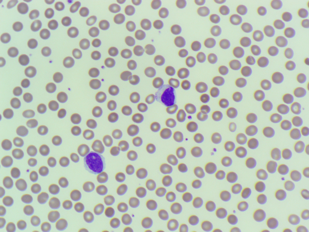
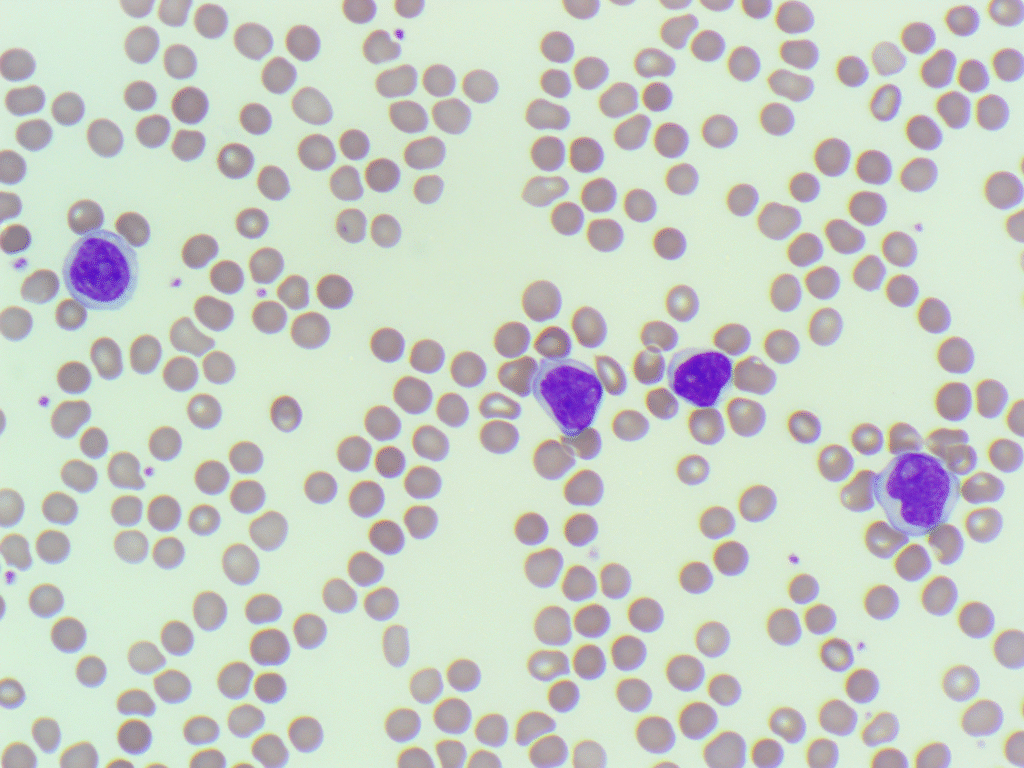
The findings are in keeping with a lymphoproliferative disorder (LPD). Given the predominance of prolymphocytes, the picture is suggestive of Prolymphocytic Leukaemia (PLL). However, this would require confirmation by flow cytometry to establish lineage and clonality.
This case highlights the importance of reviewing analyser flags and abnormal plots, especially when a grey scattergram appears, as morphology often provides the first clue to a significant haematological malignancy.
5 Comments
Good
What is meaning of Grey scatter
In this case, the grey scatter plot is due to the analyser being unable to distinguish lymphocytes and monocytes.
Would it be possible to show display the lymphocyte gallery from CellaVision? Thx you. Gerald. And were there no smudge cells?
Hi Gerald. In this particular lab we do not use Cellavision so unable to provide those images. Also you are correct, the images in the gallery are representative of the cells seen throughout the blood film.