This week’s case comes from a 59-year-old male who presented to the Emergency Department with fatigue, shortness of breath, and spontaneous bruising that had developed over several weeks. Initial blood tests revealed a markedly elevated white blood cell count, prompting urgent haematology review.
The laboratory investigations showed:
- Full blood count: WBC = 168 × 10⁹/L
- Flow cytometry: CD34– CD45+ CD117+ HLA-DR– CD13+ CD15– CD16– CD7– CD19– CD33+ CD64+ CD14– CD56– CD71+ CD123+ CD38+
- Molecular results:
- NPM1: Mutated
- FLT3 ITD: Detected
- t(8;21) and inv(16): Not detected
Blood film images from the patient are shown below:
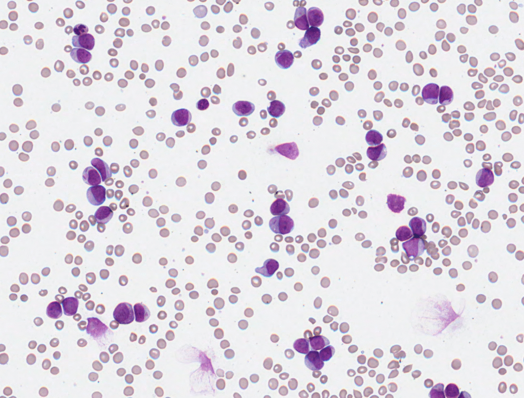
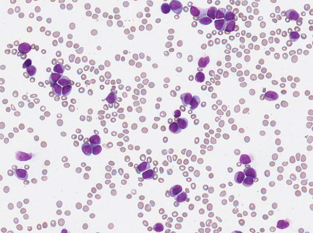
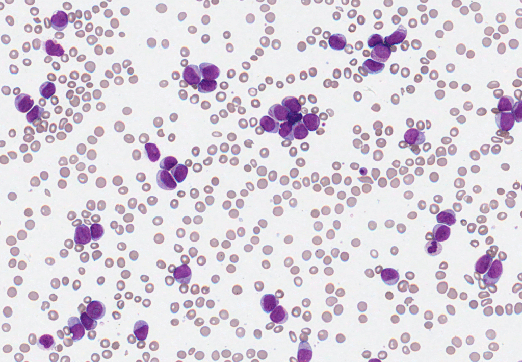
What do you make of these results, and what do you see on the blood film? Feel free to follow the discussion on LinkedIn and Twitter/X.
One Comment
Majority of large cells are blast cells with very occ showing Aur rod. Marked thrombocytopenia. Occ fragmented RBC and occ NRBC. Picture showing >20% blasts. Dx AML. Immuno shows AML probably erythroleukemia. With molecular Dx
AML with NPM1 and FLT3 ITD
Intermediate prognosis.