
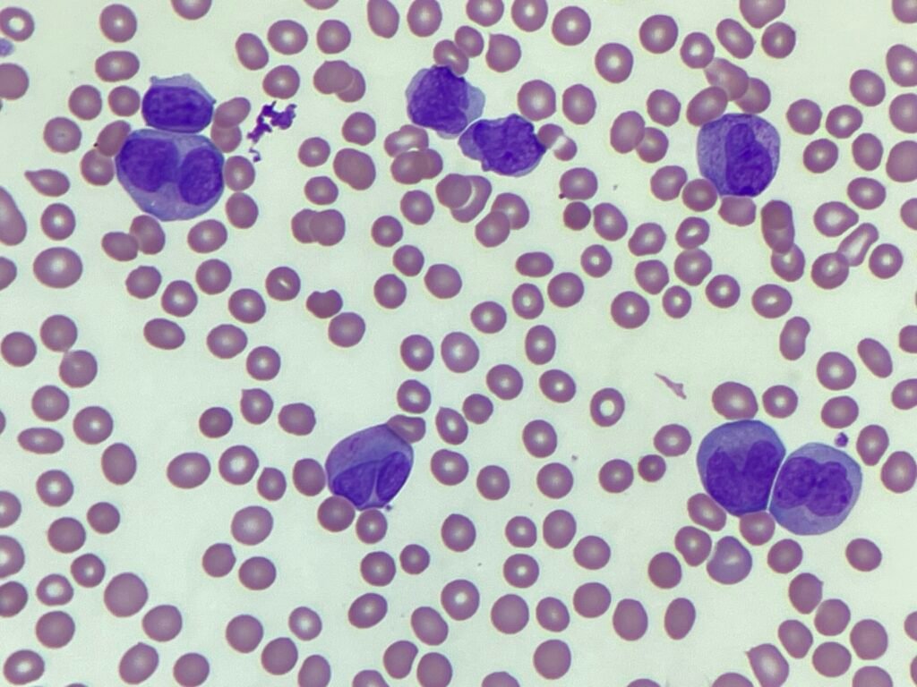
This image is from a patient whose FBC showed an increased white cell count 113.6x10⁹, haemoglobin of 78g/L and a platelet count of 58x109. The thrombocytopaenia (reduced platelets) is evident on the blood film image above. There is polychromasia and red cell anisocytosis (RDW 17.3). There are large cells with characteristic bilobed nuclei, with very occasional granules. These are seen in hypogranular or microgranular APML.
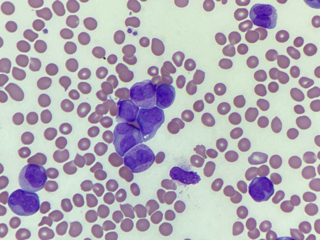
Peripheral blood film showing more bilobed or 'butterfly' cells. Though granulation is visible with light microscopy it can only be seen in some of the cells.
_____
Images: Personal photography.