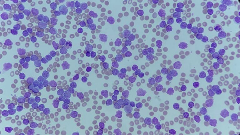
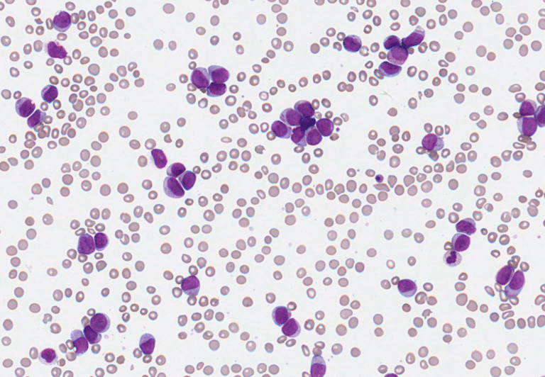
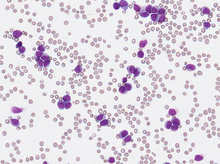
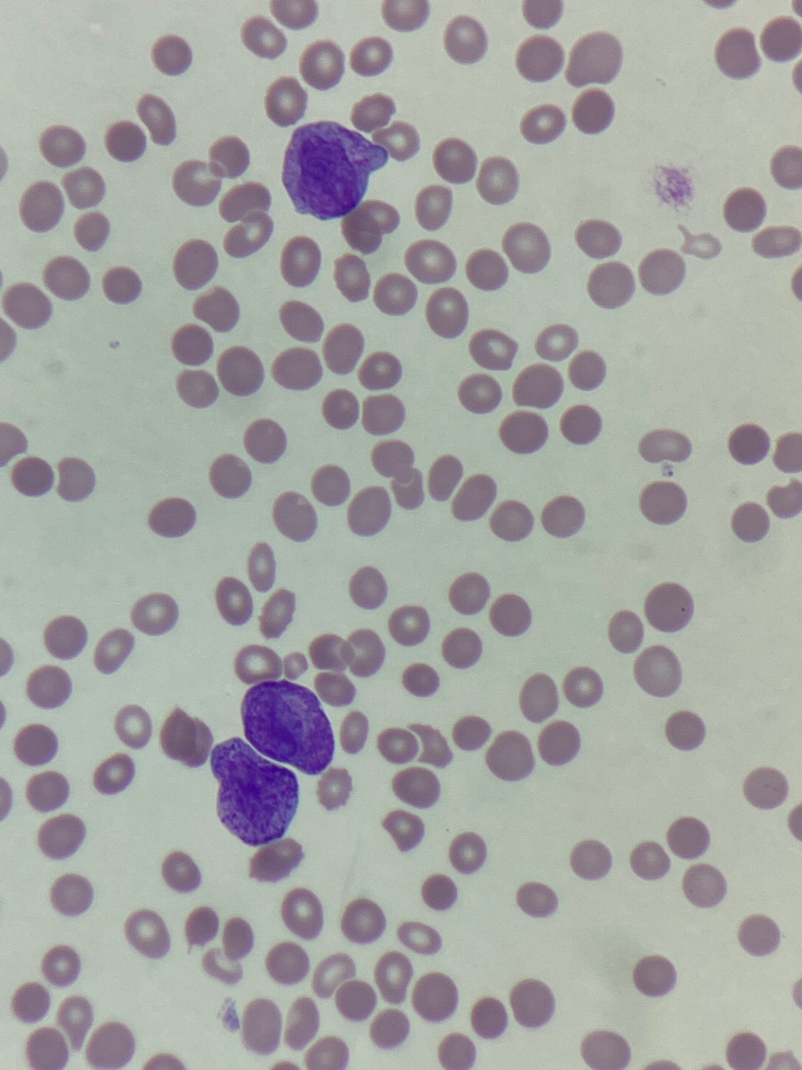
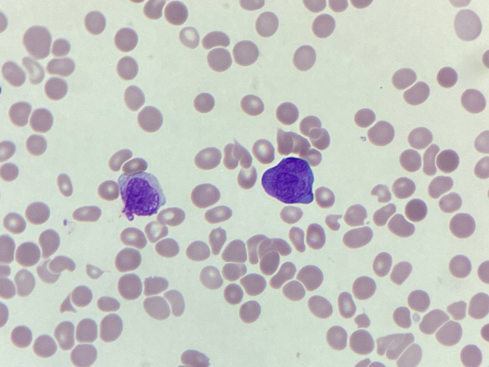
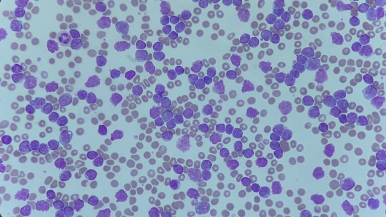
• Shop
  • Posts
  • About
  • Contact
Skip to content

🤍

£0.00 0 Cart
  • Shop
  • Posts
  • About
  • Contact
£0.00 0 Cart

🤍

white cells

MM251110: May-Hegglin anomaly
Morphology Monday | Only Cells | Platelets | White cells

MM251110: May-Hegglin anomaly

ByAnas 14/11/202517/11/2025

These features are characteristic of May–Hegglin anomaly, one of the MYH9-related disorders. This group of rare inherited conditions presents with thrombocytopenia and large platelets, sometimes accompanied by hearing loss, renal abnormalities, or cataracts.

Read More MM251110: May-Hegglin anomalyContinue

Morphology Monday | Case MM251110
Morphology Monday | Only Cells | Platelets | White cells

Morphology Monday | Case MM251110

ByAnas 10/11/202509/11/2025

A 28-year-old female was referred to haematology after routine pre-operative blood tests revealed abnormalities on the full blood count. She reports occasional easy bruising but no significant bleeding history. There’s no recent infection, and she is otherwise well.

Read More Morphology Monday | Case MM251110Continue

MM251103: Chronic myelomonocytic leukaemia
Morphology Monday | Only Cells | White cells

MM251103: Chronic myelomonocytic leukaemia

ByAnas 07/11/202503/11/2025

The combined clinical, haematological, and immunophenotypic findings are consistent with Chronic Myelomonocytic Leukaemia (CMML).

Read More MM251103: Chronic myelomonocytic leukaemiaContinue

Morphology Monday | Case MM251103
Morphology Monday | Only Cells | White cells

Morphology Monday | Case MM251103

ByAnas 03/11/202503/11/2025

This week we have a case from a 61-year-old patient who presented with persistent fatigue and unintentional weight loss. Routine bloods were performed, and the full blood count has raised some important red flags.

Read More Morphology Monday | Case MM251103Continue

MM251027: Acute myeloid leukaemia
Morphology Monday | Only Cells | White cells

MM251027: Acute myeloid leukaemia

ByAnas 31/10/202527/10/2025

The blood film showed numerous myeloid blasts with Auer rods, supporting the diagnosis of AML. Flow cytometry revealed a blast population accounting for 88.7% of total cells, with a myeloid immunophenotype

Read More MM251027: Acute myeloid leukaemiaContinue

Morphology Monday | Case MM251027
Morphology Monday | Only Cells | White cells

Morphology Monday | Case MM251027

ByAnas 27/10/202503/11/2025

This week’s case comes from a 59-year-old male who presented to the Emergency Department with fatigue, shortness of breath, and spontaneous bruising that had developed over several weeks.

Read More Morphology Monday | Case MM251027Continue

MM251013: Acute promyelocytic leukaemia (APML)
Morphology Monday | Only Cells | White cells

MM251013: Acute promyelocytic leukaemia (APML)

ByAnas 17/10/202519/10/2025

The blood film shows a genuine thrombocytopaenia, with a leukocytosis comprising predominantly of promyelocytes with Auer rods. The cells have immature nucleoli, abundant cytoplasmic granulation and approximately 25%% of the cells are bilobed. These are consistent with acute promyelocytic leukaemia.

Read More MM251013: Acute promyelocytic leukaemia (APML)Continue

Morphology Monday | Case MM251013
Morphology Monday | Only Cells | White cells

Morphology Monday | Case MM251013

ByAnas 13/10/202513/10/2025

This week’s Morphology Monday case is from a 57 year old who presented to the emergency department.

Read More Morphology Monday | Case MM251013Continue

MM251006: Lymphoproliferative disorder (?PLL)
Morphology Monday | Only Cells | White cells

MM251006: Lymphoproliferative disorder (?PLL)

ByAnas 10/10/202510/10/2025

The findings are in keeping with a lymphoproliferative disorder (LPD). Given the predominance of prolymphocytes, the picture is suggestive of Prolymphocytic Leukaemia (PLL). However, this would require confirmation by flow cytometry to establish lineage and clonality.

Read More MM251006: Lymphoproliferative disorder (?PLL)Continue

Morphology Monday | Case MM251006
Morphology Monday | Only Cells | White cells

Morphology Monday | Case MM251006

ByAnas 06/10/202506/10/2025

This week’s case comes from a 62-year-old female whose sample was flagged by the analyser, but interestingly, no differential count was produced. The WBC scattergram showed an abnormal grey plot, prompting a manual film review.

Read More Morphology Monday | Case MM251006Continue

Page navigation

Previous PagePrevious 1 2 3 Next PageNext
  • Whatsapp
  • Email
  • YouTube
  • Twitter
  • Instagram
  • Linkedin

Privacy Policy | Refund & Return Policy | Only Cells LTD © 2025