This week’s case comes from a 35-year-old patient who presented with:
- Fatigue and mild shortness of breath
- A history of intermittent anaemia
- Mild splenomegaly on examination
The laboratory results showed:
- WBC: 7.4 × 10⁹/L (normal)
- RBC: 5.69 × 10¹²/L (slightly raised)
- Hb: 102 g/L (low)
- HCT: 0.335 (low)
- MCV: 59 fL (low)
- MCH: 18.0 pg (low)
- RDW: 24.6 % (raised)
- Platelets: 157 × 10⁹/L (normal)
Special investigations:
No evidence of common haemoglobin variants (HbS, HbC) or β-thalassaemia trait. The sample was referred for further molecular analysis.
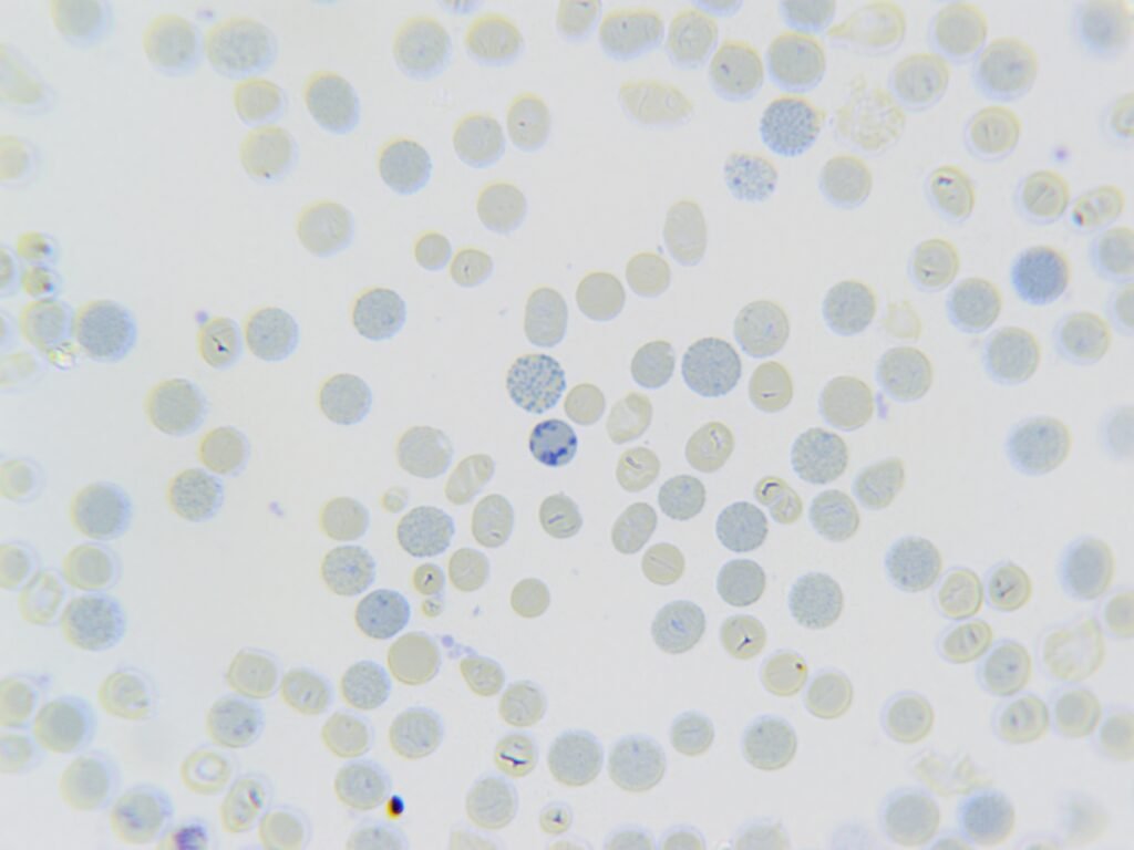
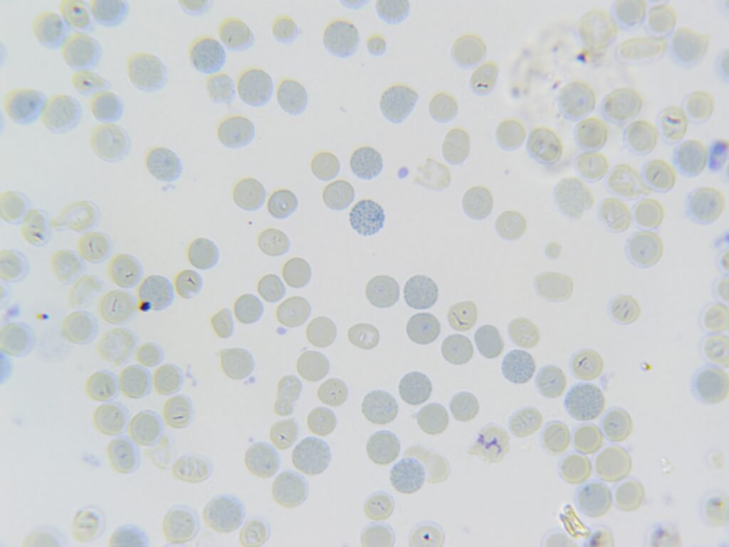
The blood film images above are stained using a special stain. What is the stain used and what is the diagnosis?
One Comment
Stain: supravital stain
Diagnosis: Hemoglobin H inclusions
Briefly explanation
What’s Hemoglobin H inclusions ?
Answer ✅
Hemoglobin H represents precipitated excess beta chains, seen only after supravital staining.
*What are the appearances?*
Answer ✅
The inclusions are small and have an even distribution that resembles the periodicity of dimples on a golf ball (hence the name “golf ball cells”).
Note
The hemoglobin H inclusions can be also seen in reticulocytes though mostly seen in matured red blood cells unfortunately they can’t be stained by Romanowsky stain so we use supravital stain