Our 72-year-old patient was referred from a neuromuscular clinic presenting with severe microcytic, hypochromic anaemia (Hb: 77g/L, MCV 56fL, MCH 16.0pg). While these indices frequently trigger an immediate assumption of severe iron deficiency, the near-normal red cell count (4.82 x 10¹²/L) and the patient’s neurological background pointed toward an entirely different pathophysiology.
The unique red cells responsible for the automated flags are acanthocytes, and their presence in a patient with neuromuscular symptoms is diagnostic of McLeod Syndrome, an X-linked recessive multi-system disorder.
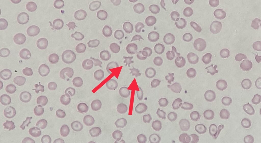
Under the microscope, the film showed prominent acanthocytes (or spur cells). These are mature red blood cells characterized by multiple, irregularly spaced, thorny or spiky cytoplasmic projections of variable length across the cell membrane.
It is vital to distinguish these from echinocytes (burr cells), which feature short, blunt, and highly symmetrical projections. The irregular remodeling seen in acanthocytes occurs because of an underlying structural imbalance within the lipid bilayer of the red cell membrane.