Thank you to everyone who commented on last week’s microcytic mystery! The combination of progressive fatigue, a high RDW, and the classic craving for ice (pica) pointed directly to the most common haematological condition encountered in the laboratory.
The 34 year old patient presented with classic symptoms of tissue hypoxia, including shortness of breath on exertion, pallor, and brittle nails. Their Full Blood Count (FBC) demonstrated a classic microcytic, hypochromic anaemia (Hb: 84 g/L, MCV: 64.2 fL, MCH: 19.5 pg) accompanied by a significant RDW (19.8%).
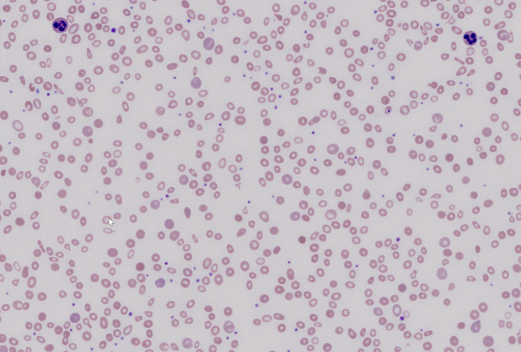
When iron stores are depleted, the bone marrow lacks the essential building blocks required to synthesise haemoglobin, leading to a highly characteristic sequential change in red cell morphology.
Under the microscope, this film provided a textbook presentation of IDA, characterised by several key features:
- Anisocytosis & Hypochromia: The red cells showed a marked variation in size (RDW), with many cells displaying a widened central pallor occupying more than the normal one-third of the cell diameter.
- Pencil Cells (Elliptocytes): The presence of occasional elongated, narrow “pencil cells” is a hallmark morphological finding highly specific to iron deficiency.
- Polychromasia: Occasional slightly larger, grayish-blue young red cells were visible, reflecting the bone marrow’s ongoing, albeit limited, attempt to release new cells into the circulation.
The elevated RDW (19.8%) is one of the most useful automated tools for distinguishing IDA from other microcytic conditions like Thalassaemia trait (where the RDW is typically normal). In IDA, as iron levels progressively decline, the bone marrow produces increasingly smaller and more poorly haemoglobinised cells over time, resulting in a highly mixed population of “normal” older cells and microcytic newer cells.