Morphology Monday | Case MM260524
This week’s case features a 72-year-old female patient whose routine FBC triggered an automatic manual film review due to some highly unusual red cell indices. The patient’s sample is from a neuromuscular clinic.
This week’s case features a 72-year-old female patient whose routine FBC triggered an automatic manual film review due to some highly unusual red cell indices. The patient’s sample is from a neuromuscular clinic.
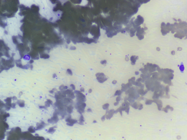
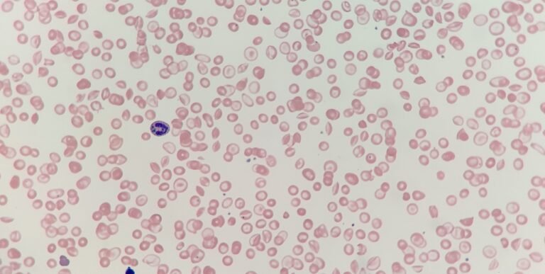
When the sample arrived in the lab, it had a “sandy” or gritty appearance inside the EDTA tube. Under the microscope, the red cells weren’t just overlapping; they were physically stuck together in irregular, massive clumps, leaving large empty spaces on the slide.
Southeast Asian ovalocytosis is a common hereditary red cell membrane disorder in parts of Southeast Asia, particularly in Malaysia, Papua New Guinea, and the Philippines. Unlike other forms of elliptocytosis, these cells are exceptionally rigid.
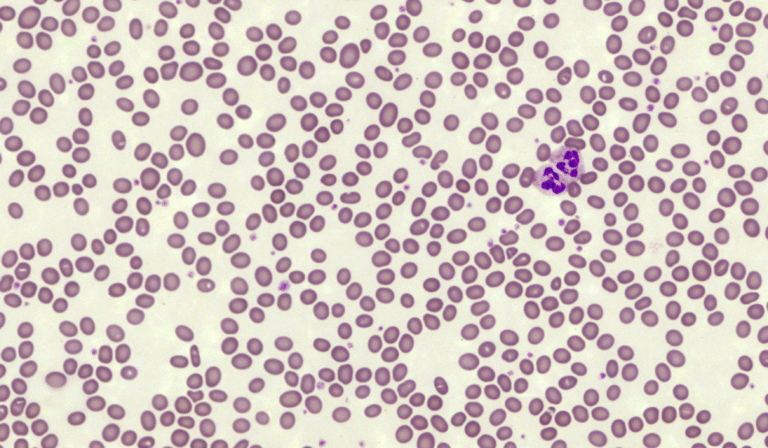
This week’s case comes from a patient of Southeast Asian descent who was recently admitted for a routine surgical workup. Interestingly, despite having no significant symptoms of anaemia, their laboratory results caught the attention of the haematology team.
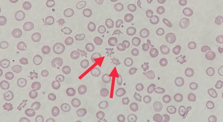
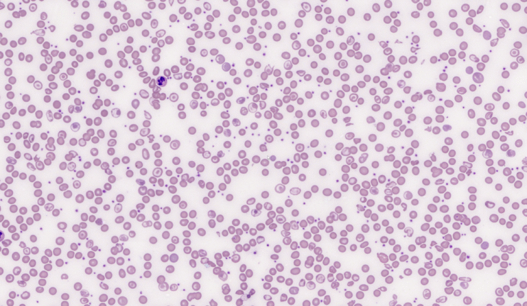
These inclusions are formally known as critical green inclusions or green neutrophilic inclusions. In clinical circles, they have earned the ominous nickname “crystals of death” due to their strong association with impending mortality.
This week’s case features a patient currently in the Intensive Care Unit (ICU) suffering from acute liver failure. The laboratory data and the resulting blood film provide a sobering look at the systemic impact of hepatic collapse.
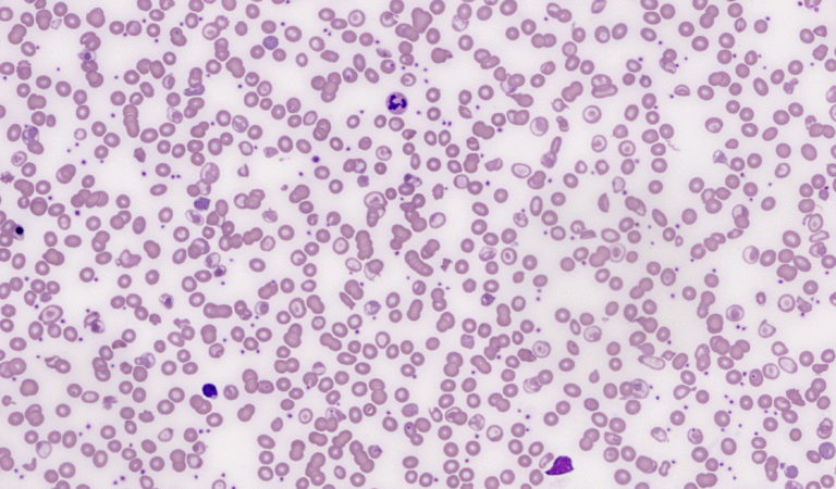
Based on these findings, the diagnosis is Haemolytic Disease of the Foetus and Newborn (HDFN) caused by Rh sensitisation, following the clinical scenario where the mother did not receive anti-D prophylaxis.
This week’s case comes from the Neonatal Intensive Care Unit. A newborn has presented with early-onset jaundice and a rapidly falling haemoglobin level.
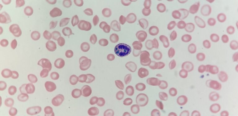
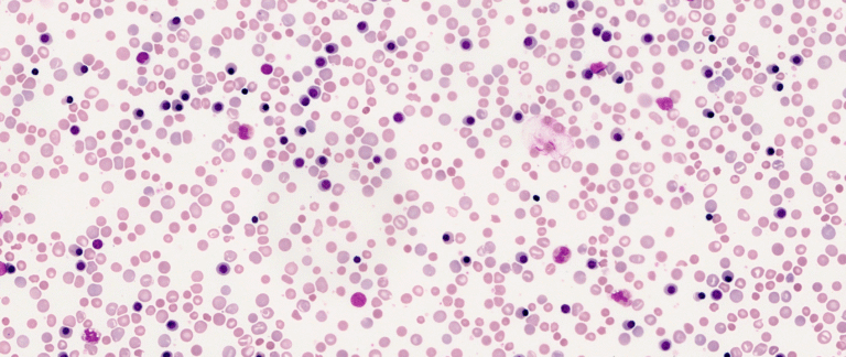
The patient is missing their spleen. In addition to the underlying HbE/β-thalassaemia, the film showed classic “post-splenectomy” features that occur when the body’s primary “quality control” filter is removed.
The blood film images are from a transfusion dependent patient who is a compound heterozygote for HbE/beta-thalassaemia.